miércoles, 30 de julio de 2014

Científicos del CNIO descubren un factor de pluripotencia embrionaria que también está activo en tejidos adultos

Fuente: http://noticias.lainformacion.com/salud/genetica/cientificos-del-cnio-descubren-un-factor-de-pluripotencia-embrionaria-tambien-esta-activo-en-tejidos-adultos_0kAoBef5eZWW8BtvzhAXH6/


Científicos del Centro Nacional de Investigaciones Oncológicas (CNIO) han descubierto que el gen NANOG, una pieza fundamental en la pluripotencia de las células madre de los embriones, regula también la división de las células adultas en los epitelios estratificados, aquellos que forman parte de la epidermis de la piel o recubren el esófago o la vagina.


Según las conclusiones del estudio, recogido en la revista 'Nature Communications', este factor podría también participar en la formación de tumores derivados de los epitelios estratificados, como los del esófago, cabeza o cuello.

Hasta la fecha se sabía que el factor de pluripotencia NANOG estaba activo durante tan solo dos días del desarrollo embrionario humano, aquellos previos a la implantación del embrión en el útero materno (entre los días 5 y 7 después de la fecundación). Durante este período crítico del desarrollo contribuye a que las células madre del embrión adquieran una amplísima capacidad de especialización que las convertirá en todos y cada uno de los tejidos que formarán el embrión.

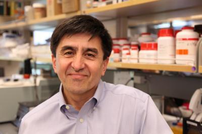
Ahora un estudio del CNIO encabezado por Manuel Serrano y Daniela Piazzolla demuestra que NANOG también tiene actividad en el organismo adulto, más allá de su función durante este breve periodo del desarrollo embrionario.

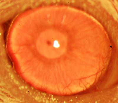
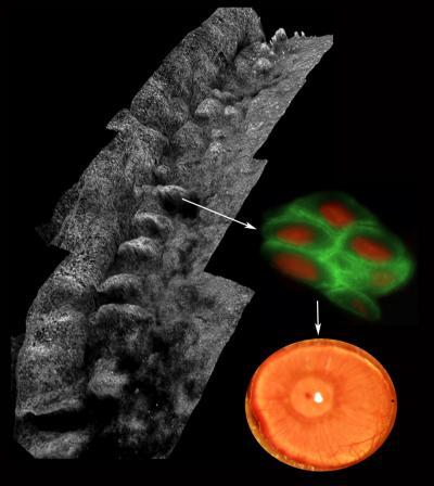
Después de analizar mediante inmunohistoquímica la presencia de NANOG en los distintos tejidos de ratón, el equipo del CNIO ha constatado que este factor está presente en epitelios adultos estratificados como los del esófago, piel o vagina.

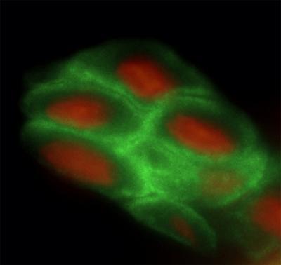
Para ello, estudiaron estirpes de ratón que permiten inducir el factor NANOG de una forma programada y limitada en el tiempo. Y según describe el artículo, el aumento de NANOG en estos ratones produce un incremento en la proliferación de las células, hiperplasia, y un aumento en el daño del ADN celular.


"Estos efectos solo se manifiestan en los epitelios estratificados, mientras que otros tejidos, como el hígado o el riñón, son indiferentes a la presencia de NANOG", ha explicado Serrano.

Estos resultados indican que hay una selectividad en los efectos de NANOG que solo se manifiestan en los epitelios estratificados. "A través de análisis globales del genoma demostramos que este factor es capaz de regular específicamente la proliferación de estos tejidos, y lo hace a través de la proteína AURKA, que está involucrada en el control de la división celular", ha añadido.

Los autores del trabajo también demuestran que NANOG está aumentado en tumores de pacientes derivados de epitelios estratificados. Además, cuando bloquearon la acción del gen mediante ARNs de interferencia el índice de proliferación de las células disminuyó.

"Esto nos indica que estas células cancerosas dependen de la actividad de NANOG para mantener su alta tasa proliferativa y sus propiedades oncológicas", según Serrano.

Stem cells may be more widespread and with greater potential than previously believed

Fuente: http://www.eurekalert.org/pub_releases/2014-06/foas-scm063014.php


With the plethora of research and published studies on stem cells over the last decade, many would say that the definition of stem cells is well established and commonly agreed upon. However, a new review article appearing in The FASEB Journal, suggests that scientists have only scratched the surface of understanding the nature, physiology and location of these cells. Specifically, the report suggests that embryonic and induced pluripotent stem cells may not be the only source from which all three germ layers in the human body (nerves, liver or heart and blood vessels) can develop. The review article suggests that adult pluripotent stem cells are located throughout the body and are able to become every tissue, provided these cells receive the right instructions.

"This study highlights the mutual role of stem cells both for regeneration and in tumor growth by featuring two sides of the same coin: stems cells in cancer and regenerative medicine," said Eckhard Alt, M.D., Ph.D., the article's lead author from the Center for Stem Cell and Developmental Biology at the University of Texas MD Anderson Cancer Center in Houston, Texas. "Our work provides novel insight on why and how nature has provided us with one universal type of stem cell that is equally distributed throughout the whole body, every organ and every tissue. Small early pluripotent stem cells are ubiquitously located in and around the blood vessels throughout the whole body and serve as a reserve army for regeneration."

In the review, Alt and colleagues suggest that small early pluripotent stem cells are able to replace any kind of tissue in the body--independent of where they comes from in the body--given that these cells receive the correct instructions. When researchers extract these cells from fat tissue, concentrated them and then injected them into diseased or injured tissue, they delivered beneficial outcomes for ailments such as heart failure, osteoarthritis, non-healing wounds, soft tissue defects, muscle, bone and tendon injuries and neurodegenerative diseases. The review also discusses how this is basically the same process that occurs in tumors, except that instead of healing or regenerating tissue, the cells work toward building a tumor. Better understanding and manipulating how these cells communicate not only will open new therapies that heal injury (heart failure, wounds, etc.), but will allow researchers to stop many cancers before they become life-threatening.

"This article suggests that the countless hours spent researching cancer and progenitor cells are finally coming to a head," said Gerald Weissmann, M.D., Editor-in-Chief of The FASEB Journal. "As the intersection between cancer and stem cell research becomes closer and clearer, all of today's medical treatments will begin to look as crude as Civil War medicine."


###

Receive monthly highlights from The FASEB Journal by e-mail. Sign up at http://www.faseb.org/fjupdate.aspx. The FASEB Journal is published by the Federation of the American Societies for Experimental Biology (FASEB). It is among the most cited biology journals worldwide according to the Institute for Scientific Information and has been recognized by the Special Libraries Association as one of the top 100 most influential biomedical journals of the past century.

FASEB is composed of 26 societies with more than 120,000 members, making it the largest coalition of biomedical research associations in the United States. Our mission is to advance health and welfare by promoting progress and education in biological and biomedical sciences through service to our member societies and collaborative advocacy.

Details: Matthias Ilmer, Jody Vykoukal, Alejandro Recio Boiles, Michael Coleman, and Eckhard Alt. Two sides of the same coin: stem cells in cancer and regenerative medicine. 
FASEB J. July 2014 28:2748-2761; doi:10.1096/fj.13-244640; 

domingo, 27 de julio de 2014

Gene discovered that activates stem cells for organ regeneration in planarians

Fuente: http://esciencenews.com/articles/2014/07/04/gene.discovered.activates.stem.cells.organ.regeneration.planarians


Researchers announced the discovery of a gene zic-1 that enables stem cells to regrow a head after decapitation in flatworm planarians. Professor Christian Petersen and Ph.D. student Constanza Vásquez-Doorman of Northwestern University discovered zic-1 by investigating planarians, an animal that uses pluripotent stem cells to regrow any missing tissue lost from injury. The study, entitled "zic-1 Expression in Planarian Neoblasts after Injury Controls Anterior Pole Regeneration," was reported in PLOS Genetics.

Many species across the animal kingdom have the ability to regenerate, but the mechanisms that connect injuries to stem cell activation and the production of new tissues are not fully understood. Developmental biologists have established that in the early embryo most animals use "tissue organizers" that secrete proteins to allow cell-cell communication for the formation of organs. But it was not clear how such tissue organizers could be produced from scratch to allow adult regeneration in animals like planarians.

A clue to the solution came from Petersen's previous research that identified the secreted protein Notum as a component of a tissue organizer needed for head regeneration in planarians. Notum drives head regeneration by inhibiting Wnt signaling, a broadly used molecular pathway used in cell-cell communication. Vásquez-Doorman and Petersen found that expression of the Notum protein and head regeneration required the zic-1 gene, which encodes a DNA-binding protein activated in stem cells early after decapitation.

Human stem cells could ultimately be used to build or repair complex tissues, but most ongoing research on these cells has focused on their ability to create individual cell types in a dish. Petersen's study identifies a potentially ancient connection between Wnt signaling and zic-1 in the use of stem cells for coordinating regenerative growth and suggests that human stem cells might someday be used to create tissue organizers for enhancement of injury repair.


Source: PLOS

Células madre en regeneración tímica

Fuente: http://www.madrimasd.org/blogs/biocienciatecnologia/2014/07/05/132993


El timo es el órgano donde se desarrollan los linfocitos T del individuo y es esencial para la generación de un Sistema Inmunitario que nos proteja frente a las infecciones y otras agresiones. Sin embargo, el timo es el primer órgano que degenera con la edad en individuos sanos, y también es sensible a tratamientos como la quimioterapia. Por lo tanto, la falta de la función de dicho órgano representa un serio problema para muchos pacientes, así como durante el envejecimiento. El trasplante de timo humano neonatal puede conducir a la reconstitución del sistema inmunológico con éxito, pero el suministro de tejido es limitado, lo que impide su uso generalizado.


La función del timo depende principalmente de un tipo de células, epiteliales tímicas, que se generan a partir de las células madre epiteliales del timo (TESC), por lo que estas últimas representan una fuente alternativa para trasplantes. Sin embargo, aún no se han desarrollado protocolos para su aislamiento y cultivo en el laboratorio.

En este sentido, el proyecto financiado por el 7º Programa Marco de la Unión Europea, THYMISTEM, coordinado por Clare Blackburn, la Universidad de Edimburgo y con la participación del grupo de María Luisa Toribio, (CBMSO; UAM-CSIC), tiene como objetivo desarrollar protocolos robustos para el cultivo a largo plazo de TESC humanas validadas funcionalmente. Se utilizarán controles de calidad estandarizados, medios optimizados y soportes adecuados para la administración de estas células a personas inmunodeprimidas, de manera que la función del timo sea completamente restaurada. El objetivo final será regenerar la función del sistema inmunológico en pacientes inmunodeprimidos.

El equipo europeo se compone, además del CSIC, de la Universidad de Edimburgo; Ruder Boskovic Institute, Croacia; the Open International University de Ucrania; Yeditepe University Vakif, Turquía; Nanopharma SA y el Sloan-Kettering Institute, en EE.UU.


Whitehead Institute researchers create 'naïve' pluripotent human embryonic stem cells

Fuente: http://www.sciencecodex.com/whitehead_institute_researchers_create_na_ve_pluripotent_human_embryonic_stem_cells-138339


For years, researchers and patients have hoped that embryonic stem cells (ESCs)—capable of forming nearly any cell type in the body—could provide insight into numerous diseases perhaps even be used to treat them. Yet progress has been hampered by the inability to transfer research and tools from mouse ESC studies to their human counterparts, in part because human ESCs are "primed" and slightly less plastic than the mouse cells.

Now Thorold Theunissen, Benjamin Powell, and Haoyi Wang, who are scientists in the lab of Whitehead Institute Founding Member Rudolf Jaenisch, have discovered how to manipulate and maintain human ESCs in a "naïve" or base pluripotent state similar to that of mouse ESCs without the use of any reprogramming factors. Their work is described in the journal Cell Stem Cell.

Naïve mouse ESCs are well-studied, and scientists have a strong understanding of how they function and mature into more specialized cells. But this understanding is of limited use in human ESC research, as the human cells look different, grow differently, and rely on different genes than mouse ESCs. According to Theunissen, the disparities between mouse and human ESCs are attributable not to species-specific differences but rather to differences of cell state.

In naïve mouse ESCs, a particular enhancer of the gene OCT4 is active, prompting the researchers to look for the presence of this marker as a means to identify rare naïve human ESCs. With this unbiased reporter system in hand, the Jaenisch team determined that a cocktail of five small molecules with a few additional growth factors can induce and support the conversion of primed human ESCs to a naïve state with or without using reprogramming factors to jumpstart the process.

By applying this cocktail to human blastocysts, the scientists could also isolate naïve human stem cells.

"This is important because if this cocktail only works in existing lines of human ESCs, you might wonder, does this really capture a distinct state or is this artificial?" says Theunissen. "Since the cocktail works directly on human blastocysts, I think it suggests that we're really capturing a cell state that is already present in the early human embryo."

Although other labs have recently reported creating naïve human ESCs, Theunissen, Powell, and Wang question these results as the cells produced through these techniques lack the gene expression and epigenetic profiles of naïve human ESCs. Yet, the Jaenisch lab believes they have now finally unlocked a way to create and maintain this important type of cell and are looking forward to exploring its potential.

"We have discovered a new pathway to generate something we believe is a totally different state of pluripotency in human ESCs that is very close to the mouse naïve state," says Jaenisch, who is also a professor of biology at MIT. "These cells may be essential for ESC technology, and that is an area we're looking forward to investigating. Now the big question for us is, does this state exist in vivo in embryos? Right now, we don't know, and that is a very interesting line of research."


NYSCF scientists 1 step closer to cell therapy for multiple sclerosis patients

Fuente: http://www.sciencecodex.com/nyscf_scientists_1_step_closer_to_cell_therapy_for_multiple_sclerosis_patients-138287


Scientists at The New York Stem Cell Foundation (NYSCF) Research Institute are one step closer to creating a viable cell replacement therapy for multiple sclerosis from a patient's own cells.

For the first time, NYSCF scientists generated induced pluripotent stem (iPS) cells lines from skin samples of patients with primary progressive multiple sclerosis and further, they developed an accelerated protocol to induce these stem cells into becoming oligodendrocytes, the myelin-forming cells of the central nervous system implicated in multiple sclerosis and many other diseases.

Existing protocols for producing oligodendrocytes had taken almost half a year to produce, limiting the ability of researchers to conduct their research. This study has cut that time approximately in half, making the ability to utilize these cells in research much more feasible.

Stem cell lines and oligodendrocytes allow researchers to "turn back the clock" and observe how multiple sclerosis develops and progresses, potentially revealing the onset of the disease at a cellular level long before any symptoms are displayed. The improved protocol for deriving oligodendrocyte cells will also provide a platform for disease modeling, drug screening, and for replacing the damaged cells in the brain with healthy cells generated using this method.

"We are so close to finding new treatments and even cures for MS. The enhanced ability to derive the cells implicated in the disease will undoubtedly accelerate research for MS and many other diseases," said Susan L. Solomon, NYSCF Chief Executive Officer.

"We hope that this protocol will be helpful to the MS field and the larger scientific community to better understand human oligodendrocyte biology and the process of myelination. This is the first step towards very exciting studies: the ability to generate human oligodendrocytes in large amounts will serve as an unprecedented tool for developing remyelinating strategies and the study of patient-specific cells may shed light on intrinsic pathogenic mechanisms that lead to progressive MS". said Dr. Valentina Fossati, NYSCF – Helmsley Investigator and senior author on the paper.

In multiple sclerosis, the protective covering of axons, called myelin, becomes damaged and lost. In this study, the scientists not only improved the protocol for making the myelin-forming cells but they showed that the oligodendrocytes derived from the skin of primary progressive patients are functional, and therefore able to form their own myelin when put into a mouse model. This is an initial step towards developing future autologous cell transplantation therapies in multiple sclerosis patients.

This important advance opens up critical new avenues of research to study multiple sclerosis and other diseases. Oligodendrocytes are implicated in many different disorders, therefore this research not only moves multiple sclerosis research forward, it allows NYSCF and other scientists the ability to study all demyelinating and central nervous system disorders.

Multiple sclerosis is a chronic, inflammatory, demyelinating disease of the central nervous system, distinguished by recurrent episodes of demyelination and the consequent neurological symptoms. Primary progressive multiple sclerosis is the most severe form of multiple sclerosis, characterized by a steady neurological decline from the onset of the disease. Currently, there are no effective treatments or cures for primary progressive multiple sclerosis and treatments relies merely on symptom management.


Descubierta una proteína que permite a las células madre del cerebro generar nuevas neuronas

Fuente: http://www.ub.edu/web/ub/es/menu_eines/noticies/2014/07/004.html


La investigadora del Instituto de Biomedicina de la Universidad de Barcelona (IBUB), Antonella Consiglio, ha participado en un trabajo que proporciona nuevas claves sobre la activación de las células madre del cerebro adulto para producir nuevas neuronas a lo largo de toda la vida. Publicada en la edición electrónica de la revista Nature Cell Biology, la investigación está coordinada por Isabel Fariñas, de la Unidad de Neurobiología Molecular de la Universidad de Valencia (UV), y en ella también colaboran los expertos Carlos López Otín (Universidad de Oviedo) y Robert Kypta (CIC bioGUNE, Bilbao).

Nuestros tejidos se renuevan constantemente gracias a las células madre, que generan nuevas células para sustituir las viejas. Estas células madre se localizan en ubicaciones muy concretas dentro de los tejidos, que se conocen como microambientes o nichos y en las que las células madre se relacionan con otros tipos de células. Aunque la regulación de estas interacciones celulares es poco conocida en general, las células madre del cerebro adulto están adheridas a otras células de su nicho por una proteína de adhesión celular, la N-cadherina, que actúa como vínculo. En este estudio, realizado en ratones, se ha demostrado que existe una proteína, la MT5-MMP, capaz de cortar ese vínculo y liberar las células madre del control del nicho.

Antonella Consiglio realizó la manipulación in vivo de la proteína N-cadherina mediante vectores lentivirales. De forma específica, la investigadora de la IBUB participó en la producción del vector lentiviral, en su validación a nivel celular y en la inyección estereotáctica en el cerebro del ratón, necesaria para llevar a cabo el estudio. «El objetivo de usar esta técnica fue poder examinar los efectos directos de la N-cadherina en vivo sin afectar a la formación del nicho de las células madre neurales adultas en etapas anteriores, cosa que se puede producir cuando se usan ratones transgénicos», explica la neurobióloga y experta en terapia génica.

«El trabajo nos permite conocer mejor las relaciones entre las células madre y su entorno, e identifica dianas moleculares sobre las que poder actuar para potenciar la activación de estas células durante la renovación de los tejidos o en procesos regenerativos», señala Eva Porlan (UV), primera autora del trabajo.

Por otro lado, la activación descontrolada de las células madre puede dar lugar a tumores. Por eso, este trabajo está vinculado a otros aspectos de la investigación orientados a comprender cómo se controla la activación normal de las células madre, con objeto de encontrar soluciones terapéuticas para la formación de tumores causados por la pérdida de dicho control. Además, este nuevo descubrimiento puede ayudar a descubrir estrategias específicas para tratar o incluso prevenir enfermedades degenerativas, como el alzheimer o el parkinson.

La investigación de Antonella Consiglio se centra en el campo de la investigación con células madre y la medicina regenerativa. Su laboratorio en el IBUB se ha especializado en el desarrollo de la transferencia de genes experimentales y el aprovechamiento de la tecnología para obtener nuevos conocimientos sobre los procesos biológicos fundamentales para la medicina molecular, como la actividad de las células madre y la neurogénesis.

Activación artificial del proceso celular de autoorganización que da forma a un embrión humano

Fuente: http://noticiasdelaciencia.com/not/10809/activacion-artificial-del-proceso-celular-de-autoorganizacion-que-da-forma-a-un-embrion-humano/es/


Aproximadamente siete días después de la concepción, sucede algo notable en el grupo de células que acabará convirtiéndose en un nuevo ser humano: Éstas empiezan a especializarse. Adoptan características que comienzan a dar pistas sobre su destino definitivo como células de la piel, del cerebro, de la musculatura o de cualquiera de los alrededor de 200 tipos de células que existen en las personas, y comienzan a formar capas definidas.


Aunque los científicos han estudiado este proceso en animales, y han intentado promover que las células madre embrionarias adopten las formas deseadas mediante la táctica de inundarlas con señales químicas incitadoras de la transformación específica, hasta ahora el proceso no ha podido ser replicado en el laboratorio. Un equipo de investigadores liderado por Ali Brivanlou, de la Universidad Rockefeller en la ciudad estadounidense de Nueva York, lo ha intentado con un planteamiento diferente, obteniendo en esta ocasión el éxito. ¿La clave para lograrlo? Resulta que el ingrediente que faltaba es de tipo geométrico, no químico…


En el útero, las células madre embrionarias humanas reciben indicaciones del tejido que las rodea para que empiecen a formar capas. Las células en el centro comienzan a formar el ectodermo, el cerebro y la piel del embrión, mientras que aquellas que migran hacia el exterior se convierten en el mesodermo y el endodermo, destinadas a convertirse en músculo y sangre, y muchos de los órganos principales, respectivamente.


El equipo de Brivanlou confinó células madre embrionarias humanas en espacios delimitados por patrones circulares diminutos, sobre placas de cristal, que habían sido tratadas químicamente para formar los “micropatrones” que evitan que las colonias se expandan fuera de un radio específico. Cuando los investigadores introdujeron señales químicas que espolearon a las células a formar capas, constataron que las colonias que estaban geométricamente confinadas de esta forma se disponían a formar endodermo, mesodermo y ectodermo, y empezaban a organizarse tal y como lo harían bajo condiciones naturales. Las células que no estaban confinadas no lo hicieron.


Patrón de crecimiento y autoorganización. Cuarenta y dos horas después de empezar a diferenciarse, las células embrionarias se están claramente segregando en endodermo (rojo), mesodermo (azul) y ectodermo (negro). (Imagen: Ali H Brivanlou, Aryeh Warmflash, Benoit Sorre, Fred Etoc, Eric D Siggia, Universidad Rockefeller)


Vigilando las vías moleculares específicas que utilizan las células humanas para comunicarse entre sí a fin de formar patrones durante ese proceso de formar capas (una vigilancia que previamente no era posible) los investigadores averiguaron también cómo ciertas señales inhibidoras específicas, generadas en respuesta a las indicaciones químicas iniciales, actúan para impedir que todas las células dentro de una colonia sigan el mismo camino de desarrollo.


Conocer a fondo lo que ocurre cuando miembros individuales de un grupo de células madre embrionarias empiezan a especializarse por vez primera y a organizarse en capas, hasta dar lugar a órganos de un nuevo ser, será decisivo para hacer realidad en toda su magnitud la promesa de la medicina regenerativa, tal como subraya Brivanlou. “Nos acerca a la posibilidad del reemplazo de órganos formados en placas de Petri, y a heridas que se cierren y curen con rapidez”.


viernes, 25 de julio de 2014

El banco familiar Crio-Cord estrena página web con el objetivo de informar sobre las células madre del cordón umbilical

Fuente: http://noticias.lainformacion.com/arte-cultura-y-espectaculos/internet/crio-cord-estrena-pagina-web-con-el-objetivo-de-informar-sobre-las-celulas-madre-del-cordon-umbilical_4HM3cqUENzUGb9mwbfDev5/


El banco familiar de conservación de células madre del cordón umbilical Crio-Cord ha estrenado una nueva página web dirigida a los padres que ofrece información sobre los usos terapéuticos actuales y el potencial de las células madre del cordón umbilical.


Crio-Cord estrena página web con el objetivo de informar sobre las células madre del cordón umbilical


Esta web también se ha comprometido con dar información detallada sobre cuál es el potencial de las células madre, sobre cuáles son sus aplicaciones y sobre cuáles son sus limitaciones, además de publicar cuáles son las áreas que se están estudiando en la actualidad.

Hoy en día, gracias al empleo de células madre se pueden tratar más de 70 enfermedades como algunos tipos de linfomas, leucemias y anemias graves, por lo que en esta web se destaca la importancia de la conservación del cordón umbilical.

Otra de las cosas que muestra esta web es la información detallada de los nuevos campos que se están estudiando como la medicina regenerativa.

Por otra parte se explica a los padres, de manera muy gráfica y sencilla cuáles son los diferentes pasos que hay que seguir para la conservación de estas valiosas células. Se pueden encontrar también testimonios de algunas de las familias que ya han confiado en Crio-Cord, que son ya más de 59.000 en España y más de 260.000 en todo el mundo (CryoSave).

Además, interactúan con los padres y con los seguidores a través de las redes sociales ('Facebook', 'Twitter' y 'Youtube') buscando divulgar la importancia de las células madre del cordón umbilical, compartiendo noticias de interés sobre el tema y resolviendo dudas.

martes, 22 de julio de 2014

Un fármaco contra las células madre que provocan cáncer y metástasis

Fuente: http://www.larazon.es/detalle_normal/noticias/6970471/salud/un-farmaco-contra-las-celulas-madre-que-provocan-cancer-y-metastasis#.Ttt1UibZ8Tj3aom


Imagen de la formación de la metástasis en un ratón 


Un equipo andaluz de investigadores, liderado por la Universidad de Granada (UGR), ha demostrado la eficacia de un nuevo fármaco frente a las células madre cancerígenas (CMC), responsables del inicio y crecimiento del cáncer, de la recaída tras la quimioterapia y de la formación de metástasis. Según informa la UGR en una nota de prensa, este medicamento, denominado bozepinib, ha resultado efectivo en un estudio realizado en ratones y el estudio se ha publicado en la prestigiosa revista Oncotarget.

Las CMC se encuentran en los tumores en muy bajo número y tienen como característica importante la formación de las metástasis en sitios diferentes al tumor original. Debido a que en condiciones normales se encuentran en estado durmiente, sin dividirse, la quimioterapia y la radioterapia convencionales actúan sobre las células del tumor más diferenciadas, que se encuentran en división, pero no son capaces de destruir estas CMC. De hecho, tras una respuesta inicial al tratamiento, muchos pacientes con cáncer tienen recaídas debido a que estas CMC no han sido destruidas. En los últimos años, la investigación en la lucha contra el cáncer se ha centrado en la búsqueda de nuevos fármacos que se dirijan selectivamente frente a estas CMC, de tal forma que si estas células son eliminadas, el tumor será destruido en su totalidad y ello dará lugar a la curación de los pacientes.

Los científicos del grupo de investigación 'Terapias avanzadas: diferenciación, regeneración y cáncer', que dirige el profesor de la UGR Juan Antonio Marchal, han colaborado con Joaquín Campos, de la Facultad de Farmacia de la UGR, y María Angel García del Hospital Universitario 'Virgen de las Nieves' de Granada, así como con las Universidades de Jaén y Miami para el desarrollo del fármaco bozepinib.


Este nuevo medicamento tiene una actividad selectiva frente a CMC de mama, colon y melanoma. "La potente actividad antitumoral de bozepinib se debe a la inhibición de la ruta de señalización de HER2, y a que dicho fármaco inhibe la invasividad y la formación de nuevos vasos en el tumor (angiogénesis)", ha explicado el profesor Juan Antonio Marchal. Además, los investigadores han demostrado también el mecanismo específico mediante el cual bozepinib actúa frente a las CMC. Este nuevo compuesto no mostró toxicidad en ratones sanos cuando se suministró por vía intraperitoneal o por vía oral, e inhibió el crecimiento tumoral y la formación de metástasis pulmonares en los ratones a los que se les indujo el tumor.

En la actualidad, los investigadores están realizando estudios de seguridad y pretenden que dicho compuesto, así como derivados del mismo, puedan pasar a ensayos clínicos con pacientes en un futuro no muy lejano.


lunes, 21 de julio de 2014

Consiguen regenerar córneas a partir de células madre humanas adultas

Fuente: http://noticias.lainformacion.com/salud/genetica/consiguen-regenerar-corneas-a-partir-de-celulas-madre-humanas-adultas_FaEWU3Tdq4D2sqiDjedvc7/


Investigadores de Boston, en Estados Unidos, han identificado una forma de potenciar el crecimiento de nuevo tejido de la córnea humana para restaurar la visión, usando una molécula llamada ABCB5, que actúa como un marcador para las células madre limbares, muy difíciles de encontrar. La investigación, publicada en 'Nature', es uno de los primeros ejemplos conocidos de construcción de un tejido de un ser humano adulto derivado de células madre.


Consiguen regenerar córneas a partir de células madre humanas adultas


Este trabajo, una colaboración entre el 'Massachusetts Eye and Ear/Schepens Eye Research Institute' (Mass. Eye and Ear), el Hospital de Niños de Boston, el Hospital Brigham y de la Mujer y el Sistema de Salud de VA Boston, todos ellos en Estados Unidos, resulta prometedor para las víctimas de quemaduras, las víctimas de lesión química y otras personas con enfermedades que dañan los ojos.

Las células madre del limbo residen en el epitelio limbal basal del ojo, o limbo, y ayudan a mantener y regenerar el tejido corneal, por lo que su pérdida por una lesión o enfermedad es una de las principales causas de ceguera. En el pasado, se han utilizado trasplantes de tejidos o células para ayudar a la regeneración de la córnea, pero no se sabía si había células madre limbares reales en los injertos, o cuántas de ellas, y los resultados no fueron consistentes.

En este estudio, los investigadores lograron utilizar anticuerpos para detectar ABCB5 en las células madre en el tejido de donantes humanos fallecidos y las utilizaron para regenerar anatómicamente córneas humanas normales y totalmente funcionales en ratones.

"Las células madre del limbo son muy raras y que el trasplante tenga éxito depende de estas células. Con este hallazgo será ahora mucho más fácil restaurar la superficie de la córnea", explica Bruce Ksander, del 'Mass. Eye and Ear', y coautor principal del estudio con su compañero postdoctoral Paraskevi Kolovou.

ABCB5 fue originalmente descubierta en el laboratorio de Markus Frank, del Hospital de Niños de Boston, y Natasha Frank, del Sistema de Salud de VA Boston y el Hospital Brigham y de la Mujer, con otros investigadores de alto nivel. En el nuevo trabajo, utilizando un modelo de ratón desarrollado por el laboratorio de Frank, vieron que ABCB5 se produce en las células madre del limbo y es necesaria para su mantenimiento y supervivencia y para el desarrollo de la córnea y su reparación, de forma que los ratones que carecen de un gen funcional ABCB5 perdieron sus poblaciones de células madre del limbo y sus córneas se regeneraron de forma pobre tras una lesión.

"ABCB5 permite que las células madre del limbo sobrevivan y las protege de la apoptosis [muerte celular programada]", subraya Markus Frank. "El modelo de ratón nos permitió entender por primera vez el papel de ABCB5 en el desarrollo normal y es muy importante para el campo de las células madre en general", añade Natasha Frank, que está trabajando con la industria biofarmacéutica para desarrollar un anticuerpo ABCB5 a nivel clínico que cumpla con los requisitos para su aprobación por las autoridades en Estados Unidos.

Researchers regrow corneas -- first known tissue grown from an adult human stem cell

Fuente: http://www.sciencecodex.com/researchers_regrow_corneas_first_known_tissue_grown_from_an_adult_human_stem_cell-136836


Boston researchers have identified a way to enhance regrowth of human corneal tissue to restore vision, using a molecule known as ABCB5 that acts as a marker for hard-to-find limbal stem cells. This work, a collaboration between the Massachusetts Eye and Ear/Schepens Eye Research Institute (Mass. Eye and Ear), Boston Children's Hospital, Brigham and Women's Hospital and the VA Boston Healthcare System, provides promise to burn victims, victims of chemical injury and others with damaging eye diseases. The research, published in Nature, is also one of the first known examples of constructing a tissue from an adult-derived human stem cell.


Limbal stem cells reside in the eye's basal limbal epithelium, or limbus, and help maintain and regenerate corneal tissue. Their loss due to injury or disease is one of the leading causes of blindness. In the past, tissue or cell transplants have been used to help the cornea regenerate, but it was unknown whether there were actual limbal stem cells in the grafts, or how many, and the outcomes were not consistent.


In this study, researchers were able to use antibodies detecting ABCB5 to zero in on the stem cells in tissue from deceased human donors and use them to regrow anatomically correct, fully functional human corneas in mice.


This is a restored functional cornea following transplantation of human ABCB5-positive limbal stem cells to limbal stem cell-deficient mice. Transplants consisting of human ABCB5-positive limbal stem cells resulted in restoration and long-term maintenance of a normal clear cornea, whereas control mice that received either no cells or ABCB5-negative cells failed to restore the cornea.
(Photo Credit: Kira Lathrop, Bruce Ksander, Markus Frank, and Natasha Frank.)


"Limbal stem cells are very rare, and successful transplants are dependent on these rare cells," says Bruce Ksander, Ph.D., of Mass. Eye and Ear, co-lead author on the study with post-doctoral fellow Paraskevi Kolovou, M.D. "This finding will now make it much easier to restore the corneal surface. It's a very good example of basic research moving quickly to a translational application."


ABCB5 was originally discovered in the lab of Markus Frank, M.D., of Boston Children's Hospital, and Natasha Frank, M.D., of the VA Boston Healthcare System and Brigham and Women's Hospital, co-senior investigators on the study, as being produced in tissue precursor cells in human skin and intestine. In the new work, using a mouse model developed by the Frank lab, they found that ABCB5 also occurs in limbal stem cells and is required for their maintenance and survival, and for corneal development and repair. Mice lacking a functional ABCB5 gene lost their populations of limbal stem cells, and their corneas healed poorly after injury.


"ABCB5 allows limbal stem cells to survive, protecting them from apoptosis [programmed cell death]," says Markus Frank. "The mouse model allowed us for the first time to understand the role of ABCB5 in normal development, and should be very important to the stem cell field in general." according to Natasha Frank.


Composite image depicting the palisades of Vogt within the human limbus (left), ABCB5-positive limbal stem cells isolated from the palisades (right; ABCB5 -- green, nucleus -- red) and a restored functional cornea following transplantation of human ABCB5-positive limbal stem cells to limbal stem cell-deficient mice (bottom right).
(Photo Credit: Kira Lathrop, Bruce Ksander, Markus Frank, and Natasha Frank.)


Markus Frank is working with biopharmaceutical industry to develop a clinical-grade ABCB5 antibody that would meet U.S. regulatory approvals. "A single lab cannot do a study like this," says Natasha Frank, also affiliated with the Harvard Stem Cell Institute. "It integrates genetics, knockout mice, antibodies, transplantation—a lot of technical expertise that we were lucky came together in a very nice way."


This is an image depicting the palisades of Vogt within the human limbus. The human limbal architecture shows the palisades of Vogt in whole mounted tissue labeled with collagen VII and imaged with a laser scanning confocal microscope. Image is a stitched Z stack series.
(Photo Credit: Kira Lathrop, Bruce Ksander, Markus Frank, and Natasha Frank.)


Nuclear transfer appears superior for creating embryonic stem cells

Fuente: http://www.sciencecodex.com/nuclear_transfer_appears_superior_for_creating_embryonic_stem_cells-136839


Scientists at Oregon Health & Science University have found that a process called "somatic cell nuclear transfer" is much better and more accurate at reprogramming human skin cells to become embryonic stem cells — capable of transforming into any cell type in the body — than an alternative process that produces cells similar to embryonic stem cells, but with many more epigenetic abnormalities.

The findings come from a team led by OHSU's Shoukhrat Mitalipov, Ph.D., with collaborators from the University of California, San Diego School of Medicine and the Salk Institute for Biological Studies. The findings, published online in the journal Nature, could have major implications for how science continues to develop better embryonic stem cells and stem cell therapies — which many hope could someday revolutionize how medicine treats and cures a wide range of diseases.

Scientists believe stem cell therapies hold the promise of replacing human cells damaged through injury or illness, meaning embryonic stem cells could someday treat or cure Parkinson's disease, multiple sclerosis, spinal cord injuries and many other diseases and conditions.

Mitalipov, director of OHSU's Center for Embryonic Cell and Gene Therapy, has been an international leader in embryonic stem cell research, especially since he and his team's breakthrough in May 2013 when they announced they had successfully used somatic cell nuclear transfer to convert human skin cells into embryonic stem cells. In 2007, Mitalipov's team successfully transformed monkey skin cells into embryonic stem cells.

The somatic cell nuclear transfer process Mitalipov and his team use involves transplanting the nucleus of one cell, containing an individual's DNA, into an egg cell that has had its genetic material removed. The unfertilized egg then develops and eventually produces stem cells.

Since 2006, however, many scientists have been focused on a different process to convert adult somatic cells — for instance, skin cells — into something very much like embryonic stem cells. The process happens through the introduction of four specific genes into the adult cell and produces transformed cells called induced pluripotent stem cells — or iPS cells. These cells, like embryonic stem cells, are capable of transforming into any type of cell in the body. The iPS process was pioneered by Japanese scientist Shinya Yamaka in 2006, who shared the 2012 Nobel Prize for his work.


Shoukhrat Mitalipov, Ph.D., Oregon Health & Science University, led a team that found that a process called "somatic cell nuclear transfer" is much better and more accurate at reprogramming human skin cells to become embryonic stem cells -- capable of transforming into any cell type in the body -- than an alternative process that produces cells similar to embryonic stem cells, but with many more epigenetic abnormalities. (Photo Credit: Oregon Health & Science University)


Since Yamaka's discovery, however, it's been known that iPS cells do not match exactly to genuine embryonic stem cells. That could affect how they can be used in stem cell research or therapies.

The current research is the first to more deeply compare the somatic cell nuclear transfer and iPS methods — and to analyze how the SCNT and iPS methods worked when they each started with the same set of skin cells.

The study found that stem cells produced from the skin cells by the SCNT method were almost identical to embryonic stem cells produced through in vitro fertilized eggs — which are considered the "gold standard." The iPS cells made from the same skin cells, in contrast, retained a large number of the skin cells' characteristics, referred to as "memory."

"We think the difference is remarkable," Mitalipov said. "The SCNT method faithfully erases the memory of the skin cell and converts it into an embryonic stem cell. The iPS method produces a cell with significant differences and abnormalities."

"If you believe that gene expression is important, which we do, then the closer you get to the gene expression patterns of embryonic stem cells, the better," said co-author Joseph R. Ecker, Ph.D., professor and director of Salk's Genomic Analysis Laboratory and co-director of The Center of Excellence for Stem Cell Genomics. "Right now, nuclear transfer cells look closer to the embryonic stem cells than do the iPS cells."

"If we can figure out how the egg manages to do a better job at reprogramming compared to the iPS method, it may help us develop a better iPS method," says co-author Louise C. Laurent, assistant professor in the Department of Reproductive Medicine at the University of California, San Diego.

Until then, Mitalipov said, he believes the SCNT method is a much better candidate for cell replacement therapies. "And we hope science can continue to develop stem cell therapies — so that somebody they can help us battle and defeat diseases that are defeating us today."


domingo, 20 de julio de 2014

La combinación de células iPS y terapia génica podría curar la retinosis pigmentaria

Fuente: http://www.abc.es/salud/noticias/20140710/abci-retinosis-pigmentaria-celulas-curacion-201407101241.html


LAB OF STEPHEN H. TSANG, M.D., PH.D./COLUMBIA UNIVERSITY MEDICAL CENTER
Imagen de una retina y de una visión sana


La retinosis pigmentaria es una de las principales causas de pérdida de visión en el mundo. Pero ahora, gracias un equipo de la Universidad de Columbia (EE.UU.) podría haber una forma de curarla. El enfoque, que se explica en la revista «Molecular Therapy», se basa en la combinación de la tecnología de células madre inducidas (iPS) pluripotentes con la terapia génica. Esta aproximación, la primera en esta línea, transforma las células de la piel en células de la retina, que posteriormente se emplean en un tipo de retinosis pigmentaria, la causada por mutaciones en el gen MFRP.

Gracias a este enfoque los investigadores dirigidos por Stephen H. Tsang han demostrado que esta forma de retinosis pigmentaria causada por mutaciones en el gen MFRP altera la proteína que aporta a las células de la retina su integridad estructural. Además, han mostrado que los efectos de dichas mutaciones pueden ser revertidos mediante la terapia génica. Según los investigadores, el enfoque podría ser utilizado para diseñar terapias personalizadas para otras formas de retinosis pigmentaria, así como otras enfermedades genéticas. Tal y como subraya Tsang, la combinación de «líneas celulares específicas para cada paciente para probar la eficacia de la terapia génica dirigida a corregir precisamente la deficiencia genética proporciona otra herramienta para avanzar en el campo de la medicina personalizada».


La retinosis pigmentaria es una enfermedad infantil pero cuyos primeros síntomas no aparecen hasta la edad adulta. Se considera que es la causa de degeneración hereditaria de la retina más frecuente. Sus primeros síntomas son: ceguera nocturna o, lo que es lo mismo, lenta capacidad para adaptarse y pérdida progresiva del campo visual hasta formar una visión en túnel. Aparece de manera silenciosa y lenta, tardándose en acudir al oftalmólogo, por término medio, 15 años desde que se inician los primeros síntomas de ceguera nocturna. Su edad de aparición oscila entre los 25-40 años. Se calcula que afecta a unas 25.000 personas en España y a más de 1,5 millones en todo el mundo.

Lo cierto es que se considera a la retinosis pigmentaria como una «gran desconocida». Hasta ahora se han identificado más de 60 genes diferentes relacionados con la RP, lo que dificulta el desarrollo de modelos para estudiar la enfermedad. Los modelos animales, aunque útiles, tienen limitaciones importantes debido a las diferencias entre especies, asegura Tsang.


LAB OF STEPHEN H. TSANG, M.D., PH.D./COLUMBIA UNIVERSITY MEDICAL CENTER
Imagen de una retina con retinosis pigmentaria y cómo afecta a la visión


Los investigadores también utilizan las células de la retina humana de los bancos de ojos para estudiar retinosis pigmentaria; sin embargo, debido a que estas células reflejan la etapa final del proceso de la enfermedad, son poco útiles para averiguar cómo se desarrolla la enfermedad. Tampoco hay modelos de cultivo de tejidos humanos de retinosis pigmentaria. Por último, las células madre embrionarias humanas podrían ser útiles en la investigación de retinosis pigmentaria, pero hay muchos aspectos éticos, legales y técnicos que resolver antes de poder ser empleadas.


Ahora el uso de las células iPS ofrece una vía de eludir todas estas limitaciones. En este caso, se pueden inducir a las propias células de la piel del paciente para que regresen a un estado más básico, lo más similar al de las células madre embrionarias. Y estas células son pluripotentes, destaca el investigador, lo que significa que se pueden transformar en células especializadas de diversos tipos.

En este estudio, el equipo de la Universidad de Columbia ha empleado la tecnología de las células IPS para transformar células de la piel obtenidas de dos pacientes con retinosis pigmentaria, cada uno con una diferente mutación MFRP en las células de la retina, para crear un modelo específico de pacientes para el estudio de la enfermedad y probar posibles terapias.

Gracias al análisis de estas células, los investigadores han visto que el principal efecto de las mutaciones MFRP es interrumpir la regulación de la actina, una proteína que compone el citoesqueleto, el andamiaje que da a la célula su integridad estructural. «Normalmente el citoesqueleto se parece a una serie de hexágonos conectados, pero si una célula pierde esta estructura, también desaparece su capacidad para funcionar». Los investigadores también encontraron que MFRP trabaja en conjunto con otro gen, CTRP5, y que es necesario un equilibrio entre los dos genes para la regulación normal de la actina.


En una segunda fase del estudio, el equipo utilizó adenovirus asociados para introducir copias normales del MFRP en las células de la retina derivadas de iPS, con el objetivo de restaurar con éxito la función de las células. Además, aplicaron la terapia génica en un grupo de ratones con retinosis con mutaciones MFRP. Y los resultados mostraron que los ratones presentaron una mejoría a largo plazo en la función visual y la restauración en la cantidad de los fotorreceptores.

«El estudio proporciona pruebas, tanto in vitro como in vivo, de que la pérdida de visión causada por mutaciones MFRP potencialmente podría ser tratada a través de una terapia génica», señala el coautor Dieter Egli.

Tsang por su parte piensa que este enfoque también se podría utilizar para estudiar otras formas de retinosis pigmentaria. «Gracias a los estudios de secuenciación del genoma sabemos que hay cientos de errores genéticos asociados con la enfermedad. Pero hasta ahora hemos tenido muy pocas formas de saber si en realidad eran los causantes de la enfermedad. Pero con este enfoque ya podemos determinar si estos genes son, de hecho, la causa de la retinosis pigmentaria, podemos comprender su función y, en última instancia, desarrollar tratamientos personalizados».

USC stem cell researcher targets the 'seeds' of breast cancer metastasis

Fuente: http://www.sciencecodex.com/usc_stem_cell_researcher_targets_the_seeds_of_breast_cancer_metastasis-137379


For breast cancer patients, the era of personalized medicine may be just around the corner, thanks to recent advances by USC Stem Cell researcher Min Yu and scientists at Massachusetts General Hospital and Harvard Medical School.

In a study in Science, Yu and her colleagues report how they isolated breast cancer cells circulating through the blood streams of six patients. Some of these deadly cancer cells are the "seeds" of metastasis, which travel to and establish secondary tumors in vital organs such as the bone, lungs, liver and brain.

Yu and her colleagues managed to expand this small number of cancer cells in the laboratory over a period of more than six months, enabling the identification of new mutations and the evaluation of drug susceptibility.


Circulating tumor cells from the blood of a breast cancer patient are shown.
(Photo Credit: Image by Maria C. Donaldson and Min Yu)


If perfected, this technique could eventually allow doctors to do the same: use cancer cells isolated from patients' blood to monitor the progression of their diseases, pre-test drugs and personalize treatment plans accordingly.

In the six estrogen receptor-positive breast cancer patients in the study, the scientists found newly acquired mutations in the estrogen receptor gene (ESR1), PIK3CA gene and fibroblast growth factor receptor gene (FGFR2), among others. They then tested either alone or in combination several anticancer drugs that might target tumor cells with these mutations and identified which ones merit further study. In particular, the drug Ganetspib — also known as STA-9090 — appeared to be effective in killing tumor cells with the ESR1 mutation.

"Metastasis is the leading cause of cancer-related death," said Yu, assistant professor in the Department of Stem Cell Biology and Regenerative Medicine at the Keck School of Medicine of USC. "By understanding the unique biology of each individual patient's cancer, we can develop targeted drug therapies to slow or even stop their diseases in their tracks."


Stem cells may ease urinary incontinence, study says

Fuente: http://medicalxpress.com/news/2014-07-stem-cells-ease-urinary-incontinence.html


For the millions of women who can't cough, sneeze or laugh without losing bladder control, researchers are testing a treatment that uses stem cells to regenerate weakened urethra muscles.


In a small pilot study, European researchers found that injecting stem cells isolated from patients' own fat tissue improved or eliminated stress incontinence in all participants within a year.

Stress incontinence affects about twice as many women as men because of pelvic floor strain from pregnancy and childbirth.

Most women who choose to treat the condition undergo a procedure that inserts surgical mesh between the urethra and vagina to reduce urine leakage, urologists said. But widening controversy over the use of surgical mesh makes the idea of stem cell treatment even more attractive.

"This is an application that makes sense because of the ease of access to the urethra, which isn't a difficult area to inject," said Dr. Timothy Boone, chairman of urology at Houston Methodist Hospital in Texas, who wasn't involved in the study.

Globally, similar research is under way on the use of stem cells to treat stress incontinence.

However, "a lot of other stem cell therapies are a lot more invasive," Boone added. "It's too soon to tell, but the hope would be that a significant number of women would benefit from this and avoid the possible complications of surgery."

The study is published online in the issue of the journal Stem Cells Translational Medicine.

Stress incontinence occurs when pelvic floor muscles supporting the bladder and urethra become too weak to prevent urine flow when pressure is placed on the abdomen. The problem can range from being a nuisance to highly debilitating.

Stem cells, which can develop into many possible cells or organs, are an area of intense study by scientists interested in managing or curing a wide array of serious health conditions.

For this study, researchers from Finland and the Netherlands treated five women suffering from stress incontinence with adipose (fat-derived) stem cells combined with bovine collagen gel—a bulking agent—and saline solution. The women either didn't want to undergo the mesh procedure, known as midurethral sling surgery, or had undergone unsuccessful mesh implants.

Prior to treatment, their stem cells were expanded in a lab for three weeks, then injected in the participants' urethras. After six months, one out of five patients displayed a negative "cough test" with a full bladder, meaning no urine leakage occurred.

At one year, the cough test produced no urine leakage in three of the five patients, and the other two were sufficiently satisfied to decide to end their treatment.

"This is a perfect condition for this type of stem cell technology," said Dr. Elizabeth Kavaler, a urologist at Lenox Hill Hospital in New York City. "You're basically regenerating a very focused, small part of the body, and it's very easy to monitor and see if it's working or not."

Boone and Kavaler said it's premature to speculate on the possible cost of stem cell treatment for urinary incontinence. But Kavaler noted that the ongoing daily costs of managing the condition—often including the purchase of incontinence pads—is "enormous" and that the cost of mesh surgery is at least several thousand dollars.

"I think [stem cell treatment] is a really great idea, and it's just the right part of the body to do this," she said.


More information: The U.S. National Library of Medicine has more about stress urinary incontinence.