Mostrando entradas con la etiqueta alzheimer. Mostrar todas las entradas

sábado, 1 de julio de 2017

Transforman células cutáneas en células cerebrales humanas

Fuente: http://www.tendencias21.net/Transforman-celulas-cutaneas-en-celulas-cerebrales-humanas_a43991.html


Un equipo internacional de científicos ha transformado células cutáneas en células cerebrales idénticas a las del tejido cerebral humano. El descubrimiento potenciará la investigación sobre el Alzheimer y de otras enfermedades degenerativas, ya que estas células están relacionadas con los procesos de inflamación cerebral propios de los trastornos neurológicos.


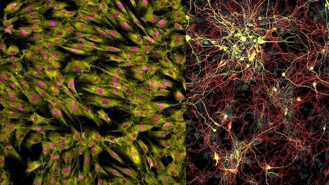
Celulas cutáneas y células cerebrales. 


Los científicos que investigan la enfermedad de Alzheimer y otras enfermedades degenerativas disponen desde ahora de la posibilidad de producir artificialmente células cerebrales, cuyo importante papel en el proceso de estas enfermedades ya ha sido demostrado. 


Un equipo internacional de científicos liderado por el Instituto de Neurología de Montreal, de la Universidad McGill, ha conseguido, con la ayuda de células cutáneas humanas, producir un tipo de células cerebrales, llamadas microgliales. 


Las microgliales son células neurogliales del tejido nervioso que forman el sistema inmunitario del sistema nervioso central. Ayudan a la conservación de las redes neuronales, así como a la respuesta del organismo a las enfermedades y las lesiones. 


El método desarrollado por estos investigadores se basa en las células madre pluripotentes inducidas (iPS), a saber, células (en este caso cutáneas) genéticamente programadas de tal manera que puedan convertirse en cualquier otro tipo de células de un organismo. 


Para transformar estas células cutáneas pluripotentes, los investigadores las han expuesto a una serie de factores de diferenciación idénticos a los que conducen a la formación de las microgliales. Las células obtenidas mediante este sistema se comportan de manera similar a como lo hacen las células microgliales. 


Con la finalidad de asegurar que las células cutáneas iniciales se transforman realmente en copias de las células microgliales, los investigadores las compararon con células cerebrales procedentes de donantes humanos. Así descubrieron que era prácticamente imposible diferenciar las células microgliales obtenidas en laboratorio de las mismas células procedentes de los tejidos humanos.


Investigaciones anteriores ya habían demostrado que las células microgliales interactúan con el péptido β-amyloïde, un ácido animado que se acumula en el cerebro de las personas que padecen la enfermedad de Alzheimer. Los péptidos son un tipo de moléculas formadas por la unión de varios aminoácidos mediante enlaces peptídicos. 


Ahora se sabe también que cuando estas células microgliales están alteradas, desencadenan una reacción inflamatoria en el cerebro, una de las causas de la aparición de la enfermedad de Alzheimer y de otros procesos degenerativos como el Parkinson o la enfermedad de Huntington. 


“Las células microgliales desempeñan un importante papel en la enfermedad de Alzheimer y otras enfermedades del sistema nervioso”, afirma Mathew Blurton-Jones, autor principal del estudio, en un comunicado de la Universidad McGill. 


“Estudios recientes han revelado que genes vinculados a un riesgo cierto de contraer la enfermedad de Alzheimer alteran el comportamiento de las células microgliales”, provocando la inflamación cerebral vinculada a la enfermedad. 


“Con la ayuda de células microgliales obtenidas en laboratorio, estamos en condiciones de comprender mejor las características biológicas de estos genes y de ensayar nuevos tratamientos”, añade Blurton-Jones. 


Por este motivo, los investigadores de este estudio, publicado en la revista Neuron, han estudiado las interacciones genéticas y físicas entre las características patológicas de la enfermedad de Alzheimer y las células microgliales obtenidas en laboratorio. 


Utilizan estas células en modelos tridimensionales del cerebro con la finalidad de comprender cómo las células microgliales interactúan con otras células cerebrales, así como su papel en la enfermedad de Alzheimer y otros trastornos neurológicos.


Referencia:


lunes, 12 de octubre de 2015

Fabrican neuronas envejecidas

Fuente: http://www.abc.es/salud/noticias/20151009/abci-celula-neurona-vejez-201510081846.html


Un nuevo método permite que las células conserven los patrones genéticos relacionados con la edad y facilita el estudio del envejecimiento.


Esta imagen muestra las diferencias entre las neuronas de las personas mayores y jóvenes cuando se convierten directamente a partir de células de la piel (izquierda) y las llamadas neuronas inducidas (derecha).


Por primera vez los científicos pueden utilizar muestras de piel de pacientes de edad avanzada para crear células cerebrales sin hacer retroceder el reloj biológico celular. La nueva técnica, que produce células parecidas a las que se encuentran en los cerebros de las personas mayores será una gran ayuda para investigar las enfermedades relacionadas con la edad, como el alzheimer y el parkinson. «Ahora podemos estudiar más fácilmente los efectos del envejecimiento en el cerebro», señala Rusty Gage, del Instituto Salk y autor principal del artículo que se publica en «Cell Stem Cell».


Según este experto, gracias a este enfoque se podrá responder a muchas preguntas acerca de la fisiología y la maquinaria molecular de las células nerviosas humanas, y no solo aquellas relacionadas con el envejecimiento saludable, «sino también con el envejecimiento patológico» añade Martin Hetzer, también del Instituto Salk.


Hasta ahora se empleaban los modelos animales, desde moscas de la fruta a ratones, para investigar las consecuencias biológicas del envejecimiento, especialmente en aquellos tejidos humanos que no se pueden obtener fácilmente en vida, como el cerebro. En los últimos años, los investigadores han recurrido cada vez más a las células madre para estudiar diversas enfermedades humanas.


Por ejemplo, se pueden obtener células de la piel de los pacientes y convertirlas en células madre pluripotentes inducidas (iPS), que tienen la capacidad de convertirse en cualquier célula en el cuerpo. A partir de ahí, los investigadores pueden programar a las células madre para convertirlas en células cerebrales. Pero este proceso, incluso cuando se obtienen células de la piel de una persona mayor, no garantiza que las células madre posean las propiedades de las células envejecidas.


Porque, señala Jerome Mertens, durante el proceso de reprogramación para crear células madre también se rejuvenecen otras propiedades de la célula, como sus firmas epigenéticas. Por eso es tan difícil investigar en el envejecimiento del cerebro humano, ya que los investigadores no han logrado verdaderas células envejecidas.


La aproximación de Gage, Hetzer y Mertens es diferente: los investigadores han convertido directamente las células de la piel en neuronas, creando lo que se llama neuronas inducidas. «Hace unos años, se demostró que es posible hacer este proceso sin pasar por el estado precursor de células madre», explica Mertens.


En esta ocasión, los investigadores han obtenido células de la piel de 19 personas, con edades desde el nacimiento hasta los 89 años, y las han transformado en células cerebrales mediante la técnica de células iPS o con el enfoque de conversión directa. A continuación, compararon las firmas epigenéticas de las neuronas resultantes con las de las células tomadas de los cerebros mediante autopsias.


Y, como se esperaba, cuando se utilizó el método de células iPS, los patrones de las neuronas eran indistinguibles entre las jóvenes y las mayores. Sin embargo, aquellas fabricadas con la técnica de conversión directa presentaban diferentes patrones de expresión de genes en función de si procedían de donantes jóvenes o de adultos mayores. «Las neuronas obtenidas mostraban diferencias en función de la edad de los donantes», asegura Mertens. Y estos patrones, añade, «en realidad muestran los cambios en la expresión génica que están implicados en el envejecimiento del cerebro».


Una vez que este método ha demostrado que retiene las firmas genéticas de la edad, esperan que pueda convertirse en una herramienta valiosa para el estudio de envejecimiento. Y, añade Cage, mientras que el trabajo actual solo probó su eficacia en la fabricación de células cerebrales, «creemos que un método similar permitirá también generar células del corazón y del hígado envejecidas».


sábado, 5 de septiembre de 2015

Logran transformar células de piel en neuronas mediante reprogramación exclusivamente química

Fuente: http://noticiasdelaciencia.com/not/15555/logran-transformar-celulas-de-piel-en-neuronas-mediante-reprogramacion-exclusivamente-quimica/es/


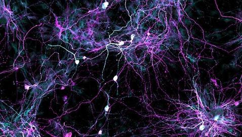
Dos laboratorios en China han tenido éxito independientemente uno del otro en la transformación de células de la piel en neuronas usando solo un “cóctel” de sustancias químicas. Uno de los dos grupos de investigación empleó células humanas de individuos sanos y de pacientes de Alzheimer. El otro grupo usó células de ratones. Los dos estudios refuerzan la idea de que una vía puramente química es una forma prometedora de avanzar en la investigación sobre la reprogramación de células, ya que permite alcanzar los mismos resultados básicos, a juzgar por lo visto, y en cambio es capaz de evitar los problemas técnicos y las preocupaciones de seguridad asociados con la estrategia más popular de reprogramación de células, la basada en el uso de factores de transcripción.


Uno de los retos de forzar a las células a cambiar de identidad es que aquellas que finalmente se obtienen como resultado del proceso pueden parecer normales pero presentar en su interior actividades diferentes a las de sus homólogas que se han formado de manera natural. Las dos investigaciones recientes proporcionan evidencias de que se pueden detectar similares expresiones genéticas, potenciales de acción, y formaciones de sinapsis tanto en las neuronas inducidas por factores de transcripción como en aquellas generadas mediante los cócteles químicos.


El estudio con células humanas lo ha realizado el equipo de la científica Jian Zhao, de los Institutos de Ciencias Biológicas de Shanghai y la Universidad Tongji también en Shanghai. Esta investigación indica además que el protocolo puramente químico puede ser utilizado para generar neuronas a partir de células cutáneas de pacientes con la enfermedad de Alzheimer.


Imagen de neuronas de ratón inducidas químicamente.


El estudio con células de ratón lo ha llevado a cabo el equipo de Hongkui Deng, del Centro de Investigación de Células Madre adscrito a la Universidad de Pekín.


miércoles, 22 de julio de 2015

Encuentran nuevos datos sobre las células madres pluripotentes

Fuente: http://www.tendencias21.net/Encuentran-nuevos-datos-sobre-las-celulas-madres-pluripotentes_a40831.html


Un equipo internacional de científicos, con la participación de la Universidad de Granada, ha aportado nuevos datos sobre las células madres pluripotentes, que podrían ayudar a entender las enfermedades inducidas por daño de los tejidos, como el Alzheimer. En concreto, han encontrado un regulador epigenético que permite que la diferenciación de las células se produzca de manera ordenada.


Células madres pluripotentes (verde) en el epiblasto (rojo) de un embrión de ratón en día 3,5 de gestación (azul). 


Un equipo internacional de científicos, en el que participa la Universidad de Granada, ha aportado nuevos datos desconocidos hasta la fecha sobre las células madre pluripotentes, una alternativa muy prometedora para el tratamiento de distintas enfermedades humanas y en especial para enfermedades inducidas por daño o degeneración de los tejidos tales como Alzheimer, Parkinson o infarto cerebral. 


Las células madre pluripotentes no pueden formar un organismo completo, pero sí cualquier otro tipo de célula correspondiente a los tres linajes embrionarios, por lo que pueden regenerar cualquier célula y tejido de un organismo. 


El trabajo, que publica la prestigiosa revista Cell Reports, revela conexiones funcionales muy importantes entre la regulación epigenética del genoma humano, las rutas de señalización celular y el fenómeno de heterogeneidad intercelular en células madre pluripotentes. 


Así, este estudio contribuye a una mejor compresión de las transiciones de linaje celular y revela aspectos desconocidos hasta ahora sobre la biología de células madre, contribuyendo así al desarrollo de distintas aplicaciones de dichas células en terapias humanas. 


David Landeira Frías, investigador del departamento de Ciencias de la Computación e Inteligencia Artificial y uno de los autores de este artículo, apunta en la nota de prensa de la universidad que recientes avances en el campo de la biomedicina “permiten reprogramar de manera artificial células del cuerpo de un organismo adulto a un estado de pluripotencia similar al encontrado durante el desarrollo embrionario temprano”.


“Una de las grandes barreras para la aplicación segura y exitosa de esta tecnología a entornos clínicos es la naturaleza heterogénea de las poblaciones de células madre; variaciones funcionales entre células de una misma población generan grandes diferencias en su comportamiento que podría conllevar el fallo de la terapia, e incluso el desarrollo de nuevas enfermedades”, advierte el investigador de la UGR. 


Las denominadas variaciones célula-célula ocurren en células con el mismo genoma y, por tanto, es muy probable que los modulares epigenéticos tengan un papel crítico en la generación de la heterogeneidad funcional. 


Mediante el uso de técnicas punteras en epigenómica, el laboratorio de David Landeira en el Centro de Genómica e Investigación Oncológica (Pfizer-Junta de Andalucía-UGR), en colaboración con el laboratorio de Amanda Fisher, del MRC-Clinical Science Centre en Reino Unido, han analizado la función de un regulador epigenético (Jarid2) en la creación de variabilidad intercelular en poblaciones de células madre pluripotentes. 


Los resultados de la investigación muestran que Jarid2 “es un factor esencial para que las células pluripotentes mantengan una interacción adecuada con las células de su entorno y puedan así llevar a cabo procesos de diferenciación celular de manera eficiente y coordinada”. 


Además, los científicos han demostrado que Jarid2 regula la heterogeneidad y función de las células pluripotentes a través de rutas de señalización tradicionalmente implicadas en diversos tipos de cáncer, por lo que el estudio también es relevante en el contexto de esta enfermedad. 


Recientemente, un equipo de científicos del Salk Institute de La Jolla (California, EEUU), en colaboración, entre otros, con investigadores del Hospital Clínic de Barcelona, la Universidad Católica de Murcia y la Clínica CEMTRO de Madrid han hallado un tipo de células madre caracterizadas por su ubicación en el embrión (específicas de una zona concreta de este) que son capaces de convertirse en cualquier tipo de tejido y que proliferan en laboratorio mejor que las otras. 


Bautizadas como “células madre pluripotentes de región selectiva” (rsPSCs, por sus siglas en inglés), las células halladas presentan unas características que hacen posible su producción a gran escala e incluso su alteración genética (editando su ADN celular). Ambas posibilidades son cruciales para las terapias de sustitución celular, y la regeneración de órganos.


Referencia bibliográfica: 

martes, 21 de julio de 2015

Células madre pluripotentes como posible terapia para alzhéimer, párkinson e infarto cerebral

Fuente: http://noticiasdelaciencia.com/not/15213/celulas-madre-pluripotentes-como-posible-terapia-para-alzheimer-parkinson-e-infarto-cerebral/


Un equipo internacional de científicos, en el que participa la Universidad de Granada (España), ha aportado nuevos datos desconocidos hasta la fecha sobre las células madre pluripotentes, una alternativa muy prometedora para el tratamiento de distintas enfermedades humanas y en especial para enfermedades inducidas por daño o degeneración de los tejidos tales como Alzheimer, Parkinson o infarto cerebral.


Su trabajo, que publica la prestigiosa revista Cell Reports, revela conexiones funcionales muy importantes entre la regulación epigenética del genoma humano, las rutas de señalización celular y el fenómeno de heterogeneidad intercelular en células madre pluripotentes.


Así, este estudio contribuye a una mejor compresión de las transiciones de linaje celular y revela aspectos desconocidos hasta ahora sobre la biología de células madre, contribuyendo así al desarrollo de distintas aplicaciones de dichas células en terapias humanas.


David Landeira Frías, investigador del departamento de Ciencias de la Computación e Inteligencia Artificial de la Universidad de Granada y uno de los autores de este artículo, apunta que recientes avances en el campo de la Biomedicina “nos permiten reprogramar de manera artificial células del cuerpo de un organismo adulto a un estado de pluripotencia similar al encontrado durante el desarrollo embrionario temprano”.


Así, estas células reprogramadas pluripotentes retienen el potencial para regenerar cualquier célula y tejido de un organismo.


“Una de las grandes barreras para la aplicación segura y exitosa de esta tecnología a entornos clínicos es la naturaleza heterogénea de las poblaciones de células madre; variaciones funcionales entre células de una misma población generan grandes diferencias en su comportamiento que podría conllevar el fallo de la terapia, e incluso el desarrollo de nuevas enfermedades”, advierte el investigador de la UGR.


Las denominadas ‘variaciones célula-célula’ ocurren en células con el mismo genoma y, por tanto, es muy probable que los modulares epigenéticos tengan un papel crítico en la generación de la heterogeneidad funcional.


Mediante el uso de técnicas punteras en epigenómica, el laboratorio de David Landeira en el Centro de Genómica e Investigación Oncológica (GENYO), Pfizer-Junta de Andalucía-UGR, en colaboración con el laboratorio de Amanda Fisher, del MRC-Clinical Science Centre en Reino Unido, han analizado la función de un regulador epigenético (Jarid2) en la creación de variabilidad intercelular en poblaciones de células madre pluripotentes.


Los resultados de la investigación muestran que Jarid2 “es un factor esencial para que las células pluripotentes mantengan una interacción adecuada con las células de su entorno y puedan así llevar a cabo procesos de diferenciación celular de manera eficiente y coordinada”.


Además, los científicos han demostrado que Jarid2 regula la heterogeneidad y función de las células pluripotentes a través de rutas de señalización tradicionalmente implicadas en diversos tipos de cáncer, por lo que el estudio también es relevante en el contexto de esta enfermedad.


jueves, 22 de enero de 2015

Research gives new hope for restoring cells in damaged brains and spinal cords

Fuente: http://medicalxpress.com/news/2015-01-cells-brains-spinal-cords.html


What motivates Penn State scientists and their students to devote countless hours trying to solve tough research mysteries? For Gong Chen, a biology professor at Penn State, the answer is rooted in a desire to help relieve the suffering of patients and their loved ones.


"I want to help people who are suffering from injuries and diseases of the brain and spinal cord," he told a crowd of Alzheimer's patients and their friends and family at Medlar Field at Lubrano Park.


During the Alzheimer's Association's Walk to End Alzheimer's Disease in October, the crowd broke into applause, cheers and some tears when Chen announced his lab's most recent research achievement.


"We have developed a revolutionary approach for reversing scarred tissues inside the brain back into normal neural tissue," he said.


Chen, who is Penn State's Verne M. Willaman Chair in the Life Sciences, directs a research team that is working on simultaneous research projects related to brain and spinal-cord disorders. These disorders include Alzheimer's disease, Parkinson's disease, amyotrophic lateral sclerosis, stroke, traumatic brain injury, spinal-cord injury, epilepsy, autism and schizophrenia.


The lab's technique for repairing scarred tissues in the brain is the latest in a recent series of discoveries that have been published by highly respected scientific journals including Cell, Nature, Cell Stem Cell and Nature Communications, among others.


The brain has neuronal cells, called neurons, and another kind called glial cells. When glial cells are healthy, they are important components of the brain's nervous system. Healthy glial cells surround neurons—the brain's nerve cells—and provide them with support, protection, insulation, oxygen and nutrients. But when neural tissue is damaged by strokes or trauma, the glial cells will react by multiplying—sometimes so excessively that they clog up the nervous system and form a glial scar, causing even more health problems for patients.


For example, glial scars that form after an injury to the spinal cord can create a blockage in the spinal cord, which may shut down the communication channel between the brain and muscles that control the legs, leaving the person unable to walk.


Recent research, published in Cell Stem Cell, describes the successful tests of Chen's lab's new technique in behavioral tests with a strain of laboratory mice known to have memory deficits and brain-cell abnormalities similar to those of human patients with Alzheimer's disease. The research demonstrated that, even in very old mice with Alzheimer's disease, Chen's team was able to regenerate many functional neurons from the internal glial cells of these mice and to replenish the lost neurons in the brains of the mice. This research raises the hope that neural-replacement therapy might someday help human patients.


Chen's lab has taken another big step by proving, as well, that human glial cells can be changed directly into neuronal cells—although this research with human brain cells can only be done in petri dishes in the lab at this time.


"Currently, there is no method available, other than the one we have developed, to repair brains by reversing glial scars back to normal neural tissues," Chen said.


The research is important because it suggests a whole new research approach for developing a novel and effective therapy for Alzheimer's disease.


"Many other research teams are injecting stem cells that are not the same as a patient's own cells into the brain to make new neurons there, but they are facing rejection by the immune system and other setbacks," Chen said. "Our technology is different. Because our method is changing the brain's own glial cells into neuronal cells, it does not require transplantation."


Chen's lab now is working on developing techniques for both gene therapy and drug therapy with the goal of moving the research through all the stages of human clinical trials that will be necessary before the therapies can be used to treat patients.


"Gene therapy will require brain surgery, but the treatments can be delivered directly into the brain. Drug therapy uses chemically-synthesized compounds and potentially can be developed into drug pills," Chen said. "It is exciting to imagine that, someday, patients may be able to take drug tablets every morning to regenerate new neurons in their brains."


Chen said he hopes that his lab's new technology eventually can be used to help patients with a range of neural injuries and diseases involving scarring of glial cells.


"This area of our research is focused on discovering effective treatments for brain disorders including Alzheimer's disease, Parkinson's disease and ALS—which involve the degeneration of neurons; stroke—which involves neuron injuries caused by events inside the brain; traumatic brain injury and spinal-cord injuries—which involve neuron damage caused by traumas from external forces like accidents and violence; and epilepsy, which can be caused by many factors," he said.


In addition to these kinds of disorders, Chen's lab also has research projects focused on autism and schizophrenia, which are neuropsychiatric disorders.


"We are working hard to move our research from the lab bench to the bedside as quickly as possible in order to directly help patients suffering from a wide range of brain and spinal-cord conditions," Chen said. "I want to let people know that our current research results can give them good reasons to hope."


jueves, 6 de noviembre de 2014

The man with a thousand brains

Fuente: http://medicalxpress.com/news/2014-10-thousand-brains.html


Forty million people worldwide are living with Alzheimer's and this is only set to increase. But tiny brains grown in culture could help scientists learn more about this mysterious disease – and test new drugs.


It sounds like something from a 1950s' B-movie: scientists growing brains in the lab. It brings to mind images of dimly lit, cobweb-filled rooms with brains pulsating in glass tanks.


The truth, of course, is far less gothic. The Wellcome Trust/Cancer Research UK Gurdon Institute, where the research is taking place, is light, airy and hi-tech. But although Dr Rick Livesey, who leads the study, does not like to call them 'mini-brains', that is in essence what they are: clusters of millions of nerve cells, electrically active and networked to each other, and no bigger than a freckle. What makes these 'brains' particularly useful is that they are diseased – they have Alzheimer's.


Almost 40 million people worldwide are living with Alzheimer's disease, and as more people live into old age, this number is set to rise. For a disease first described in 1906, surprisingly little is understood about its mechanisms, and there are no treatment options to prevent or reverse its progress. We do know, however, that the disease is characterised by the build-up in the brain of two types of protein: beta-amyloid, which clumps together into 'plaques', and tau, which accumulates in nerve cells to form 'tangles'. These proteins disrupt the behaviour of the nerve cells, which lose their connections and eventually die.


The standard way to study a disease is to use animals. Crudely speaking, you insert a human disease gene or series of genes into a mouse and observe the mechanisms that lead the animal to develop the disease. This approach is useful for asking specific questions, but doesn't show the disease process in a cohesive way. It can lead to the development of drugs that treat the disease in mice but fail when it comes to humans.


Instead, Livesey has turned to stem cells, building on research that won Sir John Gurdon, the man who gave his name to the institute at which Livesey now works, a Nobel Prize in 2012.


Most people who develop Alzheimer's begin showing symptoms in later life, from their sixties onwards. However, a small number of people – less than 1% of cases – have a genetic form of the disease that runs in their families, caused by a single change in one of three genes. If you carry the mutation, you will get Alzheimer's – and onset is typically in one's thirties or forties.


Livesey takes skin cells from patients with this familial form of Alzheimer's and reprogrammes them to become induced pluripotent stem cells, which resemble embryonic stem cells, with the ability to turn into almost any type of cell in the body. By guiding them to develop as neural stem cells, which create the brain's nerve cells, he makes two-dimensional clusters of nerve cells that model the cerebral cortex, the area of the brain that is affected by Alzheimer's. These clusters connect to each other, forming circuits – essential for modelling early stages of Alzheimer's, which affects the ability of neurons to communicate with each other. It's a very labour-intensive – and fraught – process that takes months. "We grow them in a sugar and salt mixture, and what do bacteria love but sugar and salt! You're always paranoid you're going to lose them," explained Livesey.


These clusters allow the researchers to replay a disease process that takes 30 or 40 years but over three or four months. In humans, our immune system does its best to fight off the disease, but eventually this clearance mechanism gets overwhelmed. "In the dish, these specialist clearance cells, called microglia, are absent so the disease process is faster. One of the beauties of our models is that we can add microglia and see what the effect of the immune system is and if we can use it to make things better or worse."


As well as allowing Livesey to study the fundamental biology of the disease, his mini-brains are powerful tools for screening potential drug candidates. To use animals for this purpose would mean using tens of thousands of mice which, as Livesey points out, "would be both impractical and ethically indefensible." Livesey puts the clusters into all 96 wells of a plate the size of a mobile phone and adds a different compound to each well. "We do this every two days for a month, monitoring to see if the disease gets better. We might try different doses of the same compound – we say well look, this is fine with whacking great amounts, but will it ever work as a real drug at concentrations suitable for humans?"


Thanks to a recent £2 million award from Alzheimer's Research UK, Livesey, together with colleagues at University College London, has also begun work on modelling more common forms of the disease. These share the same pathology as the familial forms, but may arise through fundamentally different mechanisms. "We know there is some overlap between the two forms, but there's a risk that we develop a treatment for familial Alzheimer's which won't work in the general population if the disease doesn't start in the same way."


The research does, however, pose an interesting ethical question: when is a model of a brain actually human? Livesey's models are made up of human cells and have many of the properties of real brains, but they cannot learn, do not think and are not sentient – they are not 'human' in that sense. "All of the nerve cells we make are excitatory – they're like 'on' switches. Real brains also have a second, inhibitory nerve cell type, which acts like an 'off' switch and modulates the neural circuits. These add an extra layer of complexity which is missing in our models.


"But honestly, we simply don't understand the cellular basis for sentience, let alone consciousness, so we probably wouldn't know it if we saw it. Nor are we clear whether it's a scale or a complexity issue. Our neural clusters are tiny, only around a million nerve cells, whereas a real brain has about 86 billion. If we made it big enough, say a kilogram, would it become a human brain? Probably not, as we're not capturing all the complexity of the system. So what if the brain model was smaller, but captured all the complexity, like a mouse brain – would that cause ethical concerns? It's a question we cannot ignore as we move forward. But we're still a long way off there yet."


miércoles, 29 de octubre de 2014

New insight on why people with Down syndrome invariably develop Alzheimer's disease

Fuente: http://www.eurekalert.org/pub_releases/2014-10/smri-nio102214.php


Amyloid plaques are found in the brains of people with Down syndrome and Alzheimer's disease.


A new study by researchers at Sanford-Burnham Medical Research Institute reveals the process that leads to changes in the brains of individuals with Down syndrome—the same changes that cause dementia in Alzheimer's patients. The findings, published in Cell Reports, have important implications for the development of treatments that can prevent damage in neuronal connectivity and brain function in Down syndrome and other neurodevelopmental and neurodegenerative conditions, including Alzheimer's disease.


Down syndrome is characterized by an extra copy of chromosome 21 and is the most common chromosome abnormality in humans. It occurs in about one per 700 babies in the United States, and is associated with a mild to moderate intellectual disability. Down syndrome is also associated with an increased risk of developing Alzheimer's disease. By the age of 40, nearly 100 percent of all individuals with Down syndrome develop the changes in the brain associated with Alzheimer's disease, and approximately 25 percent of people with Down syndrome show signs of Alzheimer's-type dementia by the age of 35, and 75 percent by age 65. As the life expectancy for people with Down syndrome has increased dramatically in recent years—from 25 in 1983 to 60 today—research aimed to understand the cause of conditions that affect their quality of life are essential.


"Our goal is to understand how the extra copy of chromosome 21 and its genes cause individuals with Down syndrome to have a greatly increased risk of developing dementia," said Huaxi Xu, Ph.D., professor in the Degenerative Diseases Program at Sanford-Burnham and senior author of the paper. "Our new study reveals how a protein called sorting nexin 27 (SNX27) regulates the generation of beta-amyloid—the main component of the detrimental amyloid plaques found in the brains of people with Down syndrome and Alzheimer's. The findings are important because they explain how beta-amyloid levels are managed in these individuals."


Xu's team found that SNX27 regulates beta-amyloid generation. Beta-amyloid is a sticky protein that's toxic to neurons. The combination of beta-amyloid and dead neurons form clumps in the brain called plaques. Brain plaques are a pathological hallmark of Alzheimer's disease and are implicated in the cause of the symptoms of dementia.


"We found that SNX27 reduces beta-amyloid generation through interactions with gamma-secretase—an enzyme that cleaves the beta-amyloid precursor protein to produce beta-amyloid," said Xin Wang, Ph.D., a postdoctoral fellow in Xu's lab and first author of the publication. "When SNX27 interacts with gamma-secretase, the enzyme becomes disabled and cannot produce beta-amyloid. Lower levels of SNX27 lead to increased levels of functional gamma-secretase that in turn lead to increased levels of beta-amyloid."


Previously, Xu and colleagues found that SNX27 deficient mice shared some characteristics with Down syndrome, and that humans with Down syndrome have significantly lower levels of SNX27. In the brain, SNX27 maintains certain receptors on the cell surface—receptors that are necessary for neurons to fire properly. When levels of SNX27 are reduced, neuron activity is impaired, causing problems with learning and memory. Importantly, the research team found that by adding new copies of the SNX27 gene to the brains of Down syndrome mice, they could repair the memory deficit in the mice.


The researchers went on to reveal how lower levels of SNX27 in Down syndrome are the result of an extra copy of an RNA molecule encoded by chromosome 21 called miRNA-155. miRNA-155 is a small piece of genetic material that doesn't code for protein, but instead influences the production of SNX27.


With the current study, researchers can piece the entire process together—the extra copy of chromosome 21 causes elevated levels of miRNA-155 that in turn lead to reduced levels of SNX27. Reduced levels of SNX27 lead to an increase in the amount of active gamma-secretase causing an increase in the production of beta-amyloid and the plaques observed in affected individuals.


"We have defined a rather complex mechanism that explains how SNX27 levels indirectly lead to beta-amyloid," said Xu. "While there may be many factors that contribute to Alzheimer's characteristics in Down syndrome, our study supports an approach of inhibiting gamma-secretase as a means to prevent the amyloid plaques in the brain found in Down syndrome and Alzheimer's."


"Our next step is to develop and implement a screening test to identify molecules that can reduce the levels of miRNA-155 and hence restore the level of SNX27, and find molecules that can enhance the interaction between SNX27 and gamma-secretase. We are working with the Conrad Prebys Center for Chemical Genomics at Sanford-Burnham to achieve this," added Xu.


###


This research was supported in part by US NIH/National Cancer Institute Grant AG038710, AG021173, NS046673, AG030197 and AG044420, and grants from the Alzheimer's Association, the Global Down Syndrome Foundation, the American Health Assistance Foundation and the National Natural Science Foundation of China.


About Sanford-Burnham Medical Research Institute

Sanford-Burnham Medical Research Institute is dedicated to discovering the fundamental molecular causes of disease and devising the innovative therapies of tomorrow. Sanford-Burnham takes a collaborative approach to medical research with major programs in cancer, neurodegeneration and stem cells, diabetes, and infectious, inflammatory, and childhood diseases. The Institute is recognized for its National Cancer Institute-designated Cancer Center, its NIH-designated Neuroscience Center Cores, and expertise in drug discovery technologies. Sanford-Burnham is a nonprofit, independent institute that employs more than 1,000 scientists and staff in San Diego (La Jolla), Calif., and Orlando (Lake Nona), Fla. For more information, visit us at sanfordburnham.org.

Sanford-Burnham can also be found on Facebook at facebook.com/sanfordburnham and on Twitter @sanfordburnham.


miércoles, 22 de octubre de 2014

Scientists sniff out unexpected role for stem cells in the brain

Fuente: http://medicalxpress.com/news/2014-10-scientists-unexpected-role-stem-cells.html


Making “scents” of new cells in the brain’s odor-processing area adult-born cells travel through the thin rostral migratory stream before settling into the olfactory bulb, the large structure in the upper right of the image.


For decades, scientists thought that neurons in the brain were born only during the early development period and could not be replenished. More recently, however, they discovered cells with the ability to divide and turn into new neurons in specific brain regions. The function of these neuroprogenitor cells remains an intense area of research. Scientists at the National Institutes of Health (NIH) report that newly formed brain cells in the mouse olfactory system—the area that processes smells—play a critical role in maintaining proper connections. The results were published in the Journal of Neuroscience.


"This is a surprising new role for brain stem cells and changes the way we view them," said Leonardo Belluscio, Ph.D., a scientist at NIH's National Institute of Neurological Disorders and Stroke (NINDS) and lead author of the study.


The olfactory bulb is located in the front of the brain and receives information directly from the nose about odors in the environment. Neurons in the olfactory bulb sort that information and relay the signals to the rest of the brain, at which point we become aware of the smells we are experiencing. Olfactory loss is often an early symptom in a variety of neurological disorders, including Alzheimer's and Parkinson's diseases.


In a process known as neurogenesis, adult-born neuroprogenitor cells are generated in the subventricular zone deep in the brain and migrate to the olfactory bulb where they assume their final positions. Once in place, they form connections with existing cells and are incorporated into the circuitry.


Dr. Belluscio, who studies the olfactory system, teamed up with Heather Cameron, Ph.D., a neurogenesis researcher at the NIH's National Institute of Mental Health, to better understand how the continuous addition of new neurons influences the circuit organization of the olfactory bulb. Using two types of specially engineered mice, they were able to specifically target and eliminate the stem cells that give rise to these new neurons in adults, while leaving other olfactory bulb cells intact. This level of specificity had not been achieved previously.


In the first set of mouse experiments, Dr. Belluscio's team first disrupted the organization of olfactory bulb circuits by temporarily plugging a nostril in the animals, to block olfactory sensory information from entering the brain. His lab previously showed that this form of sensory deprivation causes certain projections within the olfactory bulb to dramatically spread out and lose the precise pattern of connections that show under normal conditions. These studies also showed that this widespread disrupted circuitry could re-organize itself and restore its original precision once the sensory deprivation was reversed.


However, in the current study, Dr. Belluscio's lab reveals that once the nose is unblocked, if new neurons are prevented from forming and entering the olfactory bulb, the circuits remain in disarray. "We found that without the introduction of the new neurons, the system could not recover from its disrupted state," said Dr. Belluscio.


To further explore this idea, his team also eliminated the formation of adult-born neurons in mice that did not experience sensory deprivation. They found that the olfactory bulb organization began to break down, resembling the pattern seen in animals blocked from receiving sensory information from the nose. And they observed a relationship between the extent of stem cell loss and amount of circuitry disruption, indicating that a greater loss of stem cells led to a larger degree of disorganization in the olfactory bulb.


According to Dr. Belluscio, it is generally assumed that the circuits of the adult brain are quite stable and that introducing new neurons alters the existing circuitry, causing it to re-organize. "However, in this case, the circuitry appears to be inherently unstable requiring a constant supply of new neurons not only to recover its organization following disruption but also to maintain or stabilize its mature structure. It's actually quite amazing that despite the continuous replacement of cells within this olfactory bulb circuit, under normal circumstances its organization does not change," he said.


Dr. Belluscio and his colleagues speculate that new neurons in the olfactory bulb may be important to maintain or accommodate the activity-dependent changes in the system, which could help animals adapt to a constantly varying environment.


"It's very exciting to find that new neurons affect the precise connections between neurons in the olfactory bulb. Because new neurons throughout the brain share many features, it seems likely that neurogenesis in other regions, such as the hippocampus, which is involved in memory, also produce similar changes in connectivity," said Dr. Cameron.


The underlying basis of the connection between neurological disease and changes in the olfactory system is also unknown but may come from a better understanding of how the sense of smell works. "This is an exciting area of science," said Dr. Belluscio, "I believe the olfactory system is very sensitive to changes in neural activity and given its connection to other brain regions, it could lend insight into the relationship between olfactory loss and many brain disorders."


More information: "Adult Neurogenesis Is Necessary to Refine and Maintain Circuit Specificity." Journal of Neuroscience, 8 October 2014, 34(41): 13801-13810; DOI: 10.1523/JNEUROSCI.2463-14.2014


Diseñado un cultivo celular que reproduce los síntomas del alzheimer

Fuente: http://elpais.com/elpais/2014/10/13/ciencia/1413217418_488533.html


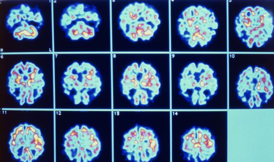
Deterioro de distintas partes del cerebro causado por el alzheimer.


Un cultivo celular que reproduce los síntomas del alzhéimer (acumulación de proteínas beta-amiloides y de ovillos de proteína tau) ha sido desarrollado por un equipo dirigido por investigadores de centros estadounidenses, liderados desde Harvard y el Instituto de Enfermedades Neurodegenerativas, ambos en Massachussets. El trabajo, que ha publicado Nature, permitirá investigar en tratamientos para la enfermedad fácilmente, al menos en las fases preclínicas.


El alzhéimer es la enfermedad neurodegenerativa más frecuente. Solo en España se calcula que la sufren unas 800.000 personas. No tiene tratamiento ni cura. Pero tampoco tiene un modelo animal adecuado. Lo que más se acerca a la enfermedad en humanos es un tipo de ratón que una variante de alzhéimer, que no reproduce en su totalidad las dos características principales de los cerebros de los afectados. Esa es la utilidad del trabajo presentado.


El proceso para conseguir una estructura similar a la de una porción de cerebro consiste en cultivar las células madre de las neuronas en un gel hasta que forman una red. La mayor parte del trabajo consiste en comprobar que estas células reproducen las características de las neuronas de los enfermos.


Este avance se une a los más avanzados para permitir un diagnóstico precoz de la enfermedad u otro más avanzado por técnicas de imagen. Con ello se puede seguir el proceso y evaluar los logros. Trabajar con el cerebro es muy difícil, y hasta hace poco el diagnóstico de alzheimer se daba por los efectos de la enfermedad en el individuo, y se confirmaba en la autopsia.


Además, los investigadores apuntan a otra posibilidad: poder estudiar si, como se cree, el proceso en las neuronas empieza por la acumulación de placas de beta-amiloides y éstas inducen la aparición de los ovillos tau. El conjunto de ambas anomalías interfiere en el funcionamiento del cerebro y causa los síntomas de la enfermedad.