Mostrando entradas con la etiqueta epilepsia. Mostrar todas las entradas

jueves, 22 de enero de 2015

Research gives new hope for restoring cells in damaged brains and spinal cords

Fuente: http://medicalxpress.com/news/2015-01-cells-brains-spinal-cords.html


What motivates Penn State scientists and their students to devote countless hours trying to solve tough research mysteries? For Gong Chen, a biology professor at Penn State, the answer is rooted in a desire to help relieve the suffering of patients and their loved ones.


"I want to help people who are suffering from injuries and diseases of the brain and spinal cord," he told a crowd of Alzheimer's patients and their friends and family at Medlar Field at Lubrano Park.


During the Alzheimer's Association's Walk to End Alzheimer's Disease in October, the crowd broke into applause, cheers and some tears when Chen announced his lab's most recent research achievement.


"We have developed a revolutionary approach for reversing scarred tissues inside the brain back into normal neural tissue," he said.


Chen, who is Penn State's Verne M. Willaman Chair in the Life Sciences, directs a research team that is working on simultaneous research projects related to brain and spinal-cord disorders. These disorders include Alzheimer's disease, Parkinson's disease, amyotrophic lateral sclerosis, stroke, traumatic brain injury, spinal-cord injury, epilepsy, autism and schizophrenia.


The lab's technique for repairing scarred tissues in the brain is the latest in a recent series of discoveries that have been published by highly respected scientific journals including Cell, Nature, Cell Stem Cell and Nature Communications, among others.


The brain has neuronal cells, called neurons, and another kind called glial cells. When glial cells are healthy, they are important components of the brain's nervous system. Healthy glial cells surround neurons—the brain's nerve cells—and provide them with support, protection, insulation, oxygen and nutrients. But when neural tissue is damaged by strokes or trauma, the glial cells will react by multiplying—sometimes so excessively that they clog up the nervous system and form a glial scar, causing even more health problems for patients.


For example, glial scars that form after an injury to the spinal cord can create a blockage in the spinal cord, which may shut down the communication channel between the brain and muscles that control the legs, leaving the person unable to walk.


Recent research, published in Cell Stem Cell, describes the successful tests of Chen's lab's new technique in behavioral tests with a strain of laboratory mice known to have memory deficits and brain-cell abnormalities similar to those of human patients with Alzheimer's disease. The research demonstrated that, even in very old mice with Alzheimer's disease, Chen's team was able to regenerate many functional neurons from the internal glial cells of these mice and to replenish the lost neurons in the brains of the mice. This research raises the hope that neural-replacement therapy might someday help human patients.


Chen's lab has taken another big step by proving, as well, that human glial cells can be changed directly into neuronal cells—although this research with human brain cells can only be done in petri dishes in the lab at this time.


"Currently, there is no method available, other than the one we have developed, to repair brains by reversing glial scars back to normal neural tissues," Chen said.


The research is important because it suggests a whole new research approach for developing a novel and effective therapy for Alzheimer's disease.


"Many other research teams are injecting stem cells that are not the same as a patient's own cells into the brain to make new neurons there, but they are facing rejection by the immune system and other setbacks," Chen said. "Our technology is different. Because our method is changing the brain's own glial cells into neuronal cells, it does not require transplantation."


Chen's lab now is working on developing techniques for both gene therapy and drug therapy with the goal of moving the research through all the stages of human clinical trials that will be necessary before the therapies can be used to treat patients.


"Gene therapy will require brain surgery, but the treatments can be delivered directly into the brain. Drug therapy uses chemically-synthesized compounds and potentially can be developed into drug pills," Chen said. "It is exciting to imagine that, someday, patients may be able to take drug tablets every morning to regenerate new neurons in their brains."


Chen said he hopes that his lab's new technology eventually can be used to help patients with a range of neural injuries and diseases involving scarring of glial cells.


"This area of our research is focused on discovering effective treatments for brain disorders including Alzheimer's disease, Parkinson's disease and ALS—which involve the degeneration of neurons; stroke—which involves neuron injuries caused by events inside the brain; traumatic brain injury and spinal-cord injuries—which involve neuron damage caused by traumas from external forces like accidents and violence; and epilepsy, which can be caused by many factors," he said.


In addition to these kinds of disorders, Chen's lab also has research projects focused on autism and schizophrenia, which are neuropsychiatric disorders.


"We are working hard to move our research from the lab bench to the bedside as quickly as possible in order to directly help patients suffering from a wide range of brain and spinal-cord conditions," Chen said. "I want to let people know that our current research results can give them good reasons to hope."


jueves, 20 de junio de 2013

UC San Francisco cell therapy raises hope for severe human forms of epilepsy

Fuente: http://www.ucsf.edu/news/2013/05/105666/epilepsy-cured-mice-using-brain-cells


Epilepsy that does not respond to drugs can be halted in adult mice by transplanting a specific type of cell into the brain, UC San Francisco researchers have discovered, raising hope that a similar treatment might work in severe forms of human epilepsy.


UCSF scientists controlled seizures in epileptic mice with a one-time transplantation of medial ganglionic eminence (MGE) cells, which inhibit signaling in overactive nerve circuits, into the hippocampus, a brain region associated with seizures, as well as with learning and memory. Other researchers had previously used different cell types in rodent cell transplantation experiments and failed to stop seizures. 


Cell therapy has become an active focus of epilepsy research, in part because current medications, even when effective, only control symptoms and not underlying causes of the disease, according to Scott C. Baraban, PhD, who holds the William K. Bowes Jr. Endowed Chair in Neuroscience Research at UCSF and led the new study. In many types of epilepsy, he said, current drugs have no therapeutic value at all.


“Our results are an encouraging step toward using inhibitory neurons for cell transplantation in adults with severe forms of epilepsy,” Baraban said. “This procedure offers the possibility of controlling seizures and rescuing cognitive deficits in these patients.”


The findings, which are the first ever to report stopping seizures in mouse models of adult human epilepsy, were published online in the journal Nature Neuroscience.


During epileptic seizures, extreme muscle contractions and often a loss of consciousness can cause seizure sufferers to lose control, fall and sometimes be seriously injured. The unseen malfunction behind these effects is the abnormal firing of many excitatory nerve cells in the brain at the same time.


In the UCSF study, the transplanted inhibitory cells quenched this synchronous, nerve-signaling firestorm, eliminating seizures in half of the treated mice and dramatically reducing the number of spontaneous seizures in the rest. Robert Hunt, PhD, a postdoctoral fellow in the Baraban lab, guided many of the key experiments.


In another encouraging step, UCSF researchers reported that they found a way to reliably generate human MGE-like cells in the laboratory, and that when transplanted into healthy mice, the cells similarly spun off functional inhibitory nerve cells. That research can be found online in the journal Cell Stem Cell.


In many forms of epilepsy, loss or malfunction of inhibitory nerve cells within the hippocampus plays a critical role. MGE cells are progenitor cells that form early within the embryo and are capable of generating mature inhibitory nerve cells called interneurons. In the Baraban-led UCSF study, the transplanted MGE cells from mouse embryos migrated and generated interneurons, in effect replacing the cells that fail in epilepsy. The new cells integrated into existing neural circuits in the mice, the researchers found.


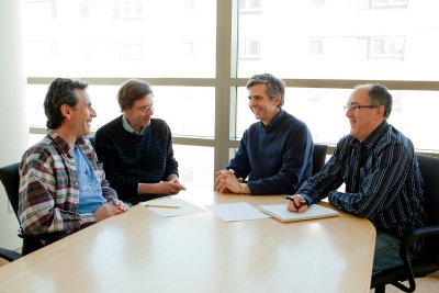
Study authors include John Rubenstein, MD, PhD; Arturo Alvarez-Buylla, PhD; and Scott C. Baraban, PhD. From left, Rubenstein, Arnold Kriegstein, MD, PhD, Alvarez-Buylla and Baraban are photographed at the Institute for Regenerative Medicine. Photo by Peter DaSilva


“These cells migrate widely and integrate into the adult brain as new inhibitory neurons,” Baraban said. “This is the first report in a mouse model of adult epilepsy in which mice that already were having seizures stopped having seizures after treatment.”


The mouse model of disease that Baraban’s lab team worked with is meant to resemble a severe and typically drug-resistant form of human epilepsy called mesial temporal lobe epilepsy, in which seizures are thought to arise in the hippocampus. In contrast to transplants into the hippocampus, transplants into the amygdala, a brain region involved in memory and emotion, failed to halt seizure activity in this same mouse model, the researchers found.


Temporal lobe epilepsy often develops in adolescence, in some cases long after a seizure episode triggered during early childhood by a high fever. A similar condition in mice can be induced with a chemical exposure, and in addition to seizures, this mouse model shares other pathological features with the human condition, such as loss of cells in the hippocampus, behavioral alterations and impaired problem solving.


In the Nature Neuroscience study, treated mice – in addition to having fewer seizures – became less abnormally agitated, less hyperactive and performed better in water-maze tests.


Additional UCSF study authors include Arturo Alvarez-Buylla, PhD, UCSF professor of neurological surgery; John Rubenstein, MD, PhD, UCSF professor of psychiatry; and Kelly Girskis, staff research associate. The research was funded by the National Institutes of Health and by the California Institute of Regenerative Medicine.

miércoles, 8 de mayo de 2013

Consiguen frenar los síntomas en ratones con epilepsia mediante un trasplante de células cerebrales

Fuente: http://www.tendencias21.net/Curan-la-epilepsia-en-ratones-con-un-trasplante-de-celulas-cerebrales_a17890.html

Hay tipos de epilepsia que no responden a fármacos, y que deben ser tratados con cirugía. Pero, desde hace un tiempo, se está investigando en la terapia celular como medio de tratamiento para estos trastornos. Científicos de EEUU han conseguido por vez primera que un trasplante de neuronas inhibitorias embrionarias sane una epilepsia en ratones. El avance aumenta la esperanza de que un tratamiento similar pueda funcionar también en las formas más graves de epilepsia humana.


La epilepsia que no responde a fármacos puede ser curada en ratones adultos mediante el trasplante de un tipo específico de célula cerebral, han constatado investigadores de la Universidad de California en San Francisco (UCSF). El hallazgo aumenta la esperanza de que un tratamiento similar pueda funcionar en las formas más graves de epilepsia humana. 

Los científicos consiguieron controlar las convulsiones epilépticas de los ratones al trasplantarles una sola vez células de la Eminencia Ganglionar Media (MGE), que es la principal fuente de interneuronas GABAérgicas de la corteza cerebral y del hipocampo. 

Estas células inhibieron la señalización de circuitos nerviosos hiperactivos presentes en el hipocampo, una región del cerebro asociada con las convulsiones, así como con el aprendizaje y la memoria. 

Previamente, ya se habían utilizado diferentes tipos de células en experimentos de trasplantes celulares con roedores, pero en estos casos no se habían conseguido detener las convulsiones. 

La terapia celular se ha convertido en un foco activo de investigación en relación con la epilepsia, en parte porque los medicamentos actuales, aun cuando a veces pueden controlar los síntomas de esta enfermedad, en algunos casos no resultan eficaces, explica Scott C. Baraban, director del estudio en un comunicado de la UCSF emitido vía Newswise. 


"Nuestros resultados son un avance alentador hacia el uso de neuronas inhibitorias para el trasplante celular en adultos con formas graves de epilepsia", afirma Baraban. "Este procedimiento ofrece la posibilidad de controlar las crisis y de recuperar los déficits cognitivos sufridos por los pacientes”. Un artículo publicado por Nature Neuroscience describe el logro al detalle. 

Durante las crisis epilépticas, se producen contracciones musculares extremas y, a menudo, una pérdida de conciencia que puede hacer que los pacientes pierdan el control y caigan, en ocasiones sufriendo lesiones graves. 

Estos síntomas tienen su origen en una activación anómala de muchas células nerviosas excitatorias del cerebro al mismo tiempo. En el estudio de la UCSF, las células inhibitorias trasplantadas desactivaron esta sincronía celular excesiva, eliminando así las convulsiones epilépticas de los ratones tratados.


El pasado dos de mayo, los científicos informaron además de que habían descubierto la manera de crear en laboratorio células MGE fiables y similares a las de los humanos, y que, cuando estas fueron trasplantadas a ratones sanos, mostraron ser células nerviosas inhibitorias funcionales. De este paso anterior se hizo eco la revista Cell Stem Cell. 


En muchas formas de epilepsia, desempeña un papel crítico la pérdida o el mal funcionamiento de las células nerviosas inhibitorias del hipocampo. 

Las células MGE son células progenitoras que se forman en el embrión y que pueden generar unas células nerviosas maduras llamadas interneuronas inhibitorias. En el estudio dirigido por Baraban, las células MGE -de embriones de ratón- trasplantadas emigraron y generaron interneuronas, remplazando las células cuyo fallo provoca la epilepsia. 

Las nuevas células se integraron en los circuitos neuronales ya existentes en los ratones, constataron los investigadores. "Estas células migran mucho y se integran en el cerebro adulto como nuevas neuronas inhibidoras", afirma Baraban. "Este es el primer registro, en modelos de ratón y de epilepsia en adultos, de ratones con convulsiones que dejaron de tenerlas tras recibir el tratamiento", añade el científico. 


El tipo de epilepsia tratada fue la epilepsia del lóbulo temporal mesial, que es resistente a los medicamentos y se suele tratar con neurocirugía. Las convulsiones producidas por este trastorno tienen su origen en el hipocampo. 

Los científicos probaron también con un trasplante de células MEG en otra parte del cerebro, la amígdala –involucrada en la memoria y la emoción- pero de este modo no consiguieron detener la actividad convulsiva de los animales. 

La epilepsia del lóbulo temporal se desarrolla a menudo en la adolescencia, en algunos casos, mucho después de un episodio de convulsiones sufrido durante la primera infancia por fiebre alta. En ratones, se puede inducir una condición similar mediante una exposición química. 

Los trasplantes hicieron que, además de tener un menor número de convulsiones, los ratones tratados estuvieran menos agitados e hiperactivos, y que obtuvieran mejores resultados en una prueba cognitiva, basada en un laberinto de agua.


Referencias bibliográficas: 

Robert F Hunt, Kelly M Girskis, John L Rubenstein, Arturo Alvarez-Buylla, Scott C Baraban. GABA progenitors grafted into the adult epileptic brain control seizures and abnormal behavior. Nature Neuroscience (2013). DOI: 10.1038/nn.3392. 

Cory R. Nicholas, Jiadong Chen, Yunshuo Tang, Derek G. Southwell, Nadine Chalmers, Daniel Vogt, Christine M. Arnold, Ying-Jiun J. Chen, Edouard G. Stanley, Andrew G. Elefanty, Yoshiki Sasai, Arturo Alvarez-Buylla, John L.R. Rubenstein, Arnold R. Kriegstein. Functional Maturation of hPSC-Derived Forebrain Interneurons Requires an Extended Timeline and Mimics Human Neural Development. Cell Stem Cell (2013). DOI:10.1016/j.stem.2013.04.005.