Noticias científicas, asesoramiento y ayuda en todo lo referente al mundo de las células madre, su conservación y aplicación.
La información contenida en este blog se ofrece solamente con carácter formativo y educativo, y no pretende sustituir las opiniones, consejos y recomendaciones de un profesional sanitario.
Las decisiones relativas a la salud deben ser tomadas por un profesional sanitario, considerando las características únicas del paciente.
Un trabajo muestra en ratones cómo detener a las células madre que originan nuevos tumores en otros órganos.
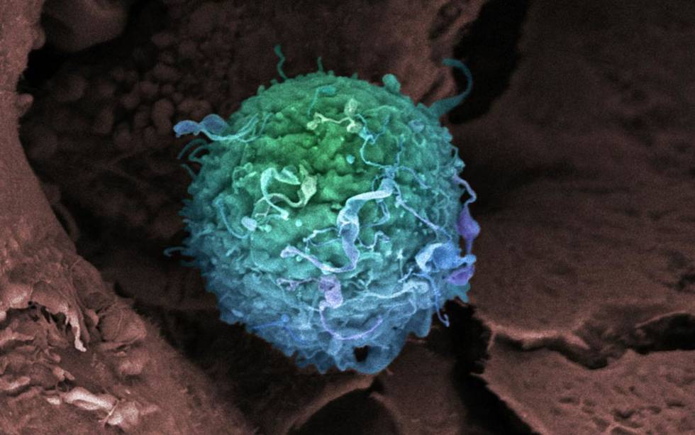
Como en muchos otros tumores, la metástasis es la principal causa de muerte en las mujeres que desarrollan cáncer de mama. Una reducida población de células dentro del tumor son capaces de sobrevivir al tratamiento y, meses o incluso años después, generar un nuevo cáncer. Las responsables de estas recaídas son las células madre del cáncer y frenarlas aportaría una de las mayores victorias imaginables contra esta enfermedad.
Un nuevo estudio publicado acerca un paso ese posible triunfo al desvelar el mecanismo que permite a las células madre del cáncer escapar al sistema inmune y generar tumores en otros órganos. “Las células madre cancerosas utilizan programas genéticos propios de las células madre normales para adquirir propiedades extra”, resume Toni Celià-Terrassa, investigador en la Universidad de Princeton (EE UU) y primer autor del estudio, publicado en Nature Cell Biology. "Eso les da capacidad de adaptación para ser más agresivas, por eso son las responsables de iniciar el crecimiento tumoral, la metástasis y la resistencia a la quimioterapia”, resalta este biólogo molecular mallorquín, que lleva cuatro años en EE UU como investigador postdoctoral.
Su trabajo muestra que tanto las células madre de la mama como las cancerosas producen una molécula de ARN que bloquea la producción de una proteína llamada LCOR. Esa proteína es fundamental para que las células madre no sean eliminadas por los interferones, unas proteínas del sistema inmune. Este mecanismo permite que las células tumorales sobrevivan sin ser vistas. El estudio ha demostrado que las pacientes con tumores triple negativo, los más agresivos y difíciles de tratar, tienen altos niveles del ARN mencionado. En sus experimentos con ratones avatar, que desarrollan tumores extraídos de las pacientes, los científicos han demostrado cómo este ARN aumenta la capacidad de las células madre para formar tumores y cómo esta se puede frenar aumentando los niveles de la proteína LCOR.
Según Celià-Terrassa, este descubrimiento podría mejorar la efectividad de la inmunoterapia, una nueva línea de tratamiento contra varios tipos de cáncer. El investigador añade que, en un futuro, se podrían desarrollar terapias combinadas con interferón y pequeñas moléculas de ARN para conseguir eliminar a esa reducida población de células dentro del tumor que son las responsables de la metástasis.
El trabajo tiene “claras implicaciones terapéuticas para atacar a las células madre del cáncer con moduladores del sistema inmune”, ha dicho Michael Clarke, investigador de la Universidad de Stanford y codescubridor de este tipo de células, en una nota de prensa emitida por Princeton. Roger Gomis, jefe del grupo de metástasis del Instituto de Investigación Biomédica, en Barcelona, opina que el trabajo aporta “un conocimiento fundamental”, aunque añade que será un reto importante poder traducirlo a posibles tratamientos a corto plazo.
Hasta el 90% de las muertes por cáncer se deben a las metástasis, pero no hay tratamientos contra ellas. Ahora, un estudio ha identificado las células que las inician en varios tipos de tumores. Para su sorpresa, los investigadores han visto que las implicadas dependen de grasas como los aceites de palma o de coco, presentes en las comidas procesadas.
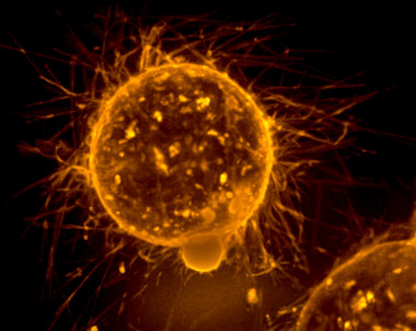
Microtentáculos en la superficie de un tumor de mama.
El cáncer es un enemigo a distancia. Nace y crece en un lugar concreto, pero hasta el 90% de las muertes que provoca dependen de los viajes que sus células emprenden por otros rincones del organismo: las metástasis.
Pero las claves de esas metástasis siguen jugando al escondite en los laboratorios. Aunque había sospechas, ni siquiera se conocían qué células concretas del tumor las iniciaban. Y, lo más importante, no existe ningún tratamiento específico ni eficaz contra ellas.
Ahora, una investigación liderada por el grupo de Salvador Aznar, investigador ICREA en el Instituto de Investigación Biomédica (IRB Barcelona), apunta a una pequeña gran revolución. El estudio que publica su equipo en la revista Nature ha dado tres saltos: identifica las células de origen de la metástasis en varios tipos de tumores; revela que se diferencian del resto porque dependen de las grasas –entre ellas, del ácido palmítico de muchas comidas procesadas–; y da con un marcador que, al inutilizarlo en ratones, previene por completo la formación de metástasis y reduce las ya existentes.
Este ha sido su viaje y hasta dónde han llegado.
“Nosotros no pretendíamos estudiar las metástasis”, comenta Salvador Aznar. “Yo llevaba tiempo analizando las células madre del cáncer y quería profundizar en un tipo particular, las células quiescentes o dormidas”.
Una teoría cada vez más aceptada sostiene que un tumor, igual que un tejido normal, tiene unas células madre que lo originan y a partir de las cuales se derivan todas las demás. Pero dentro de ellas hay un subgrupo compuesto por células madre dormidas, que parecen funcionar como una reserva. "Podrían ser más resistentes a los tratamientos y estar detrás de las recaídas tras la quimioterapia”, añade Aznar.
La sorpresa llegó cuando trataron de separarlas y aislarlas. Usando muestras de pacientes con cáncer oral vieron que había, en efecto, células madre de ciclo más lento. “Pero cuando las analizamos, ellas mismas nos dijeron que tenían algo que ver con las metástasis, porque expresaban muchos genes relacionados con éstas. Parecían adipocitos, células de la grasa. En ese momento cambiamos completamente el objetivo de la investigación”.
Para poder jugar con las células se necesita encontrar una cerradura que las diferencie. De entre todos los genes especialmente activos de las células dormidas había uno que llamaba la atención. Se llama CD36 y la proteína a la que da lugar es una puerta de entrada a las células.
Jugando con ella en ratones, los resultados fueron impactantes: cuando se aumentaba en los tumores orales, que suelen dar metástasis a los ganglios linfáticos en un 20% de las ocasiones, el porcentaje aumentaba hasta un 80% y los ganglios eran 40 veces más grandes. Al contrario, cuando se usaban anticuerpos que la bloqueaban, las metástasis disminuían entre un 80% y un 90%. En algunos casos incluso desaparecían. Y si se administraban antes de introducir las células cancerígenas, prevenían por completo su aparición. Todo ello, curiosamente, sin afectar apenas al tumor de origen.
Al revisar datos de estudios previos, el grupo de Aznar observó que el aumento de CD36 en pacientes con cáncer de pulmón, mama o vejiga también estaba relacionado con un peor pronóstico. Y cuando probaron en los ratones con melanoma o cáncer de mama, los resultados fueron muy parecidos. En el artículo hablan de un mecanismo general de metástasis.
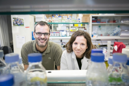
Aznar y Pascual en su laboratorio del IRB.
¿Por qué nadie lo había estudiado antes? “No lo sé”, reconoce Aznar. “Los datos estaban ahí y algunos ya habían mostrado la relación con el pronóstico, pero quizá la gente prestaba atención a aquello que le interesaba. Se parece a lo que ha sucedido con CRISPR, cuya existencia descubrió un español hace años pero hasta mucho después no hubo quien vio las posibilidades y desarrolló la técnica”.
Ahora están trabajando con MRC Technology, del Reino Unido, para desarrollar anticuerpos que puedan probarse en humanos. Ese es el verdadero salto. “Esperamos conseguirlos –comenta Aznar– y que no se queden en el camino. En cualquier caso habrá que esperar unos años”. Un aspecto positivo es que, al menos en ratones y administrados durante periodos no demasiado largos de tiempo, los efectos secundarios no parecen graves. Otro: los ratones no parecían generar resistencias, presentes en la inmensa mayoría de las terapias dirigidas contra el cáncer.
Para Joan Massagué -asesor científico del propio IRB y director del Instituto Sloan Kettering-, que no ha participado en el estudio, “este trabajo es una excelente contribución al creciente conocimiento sobre las células que originan las metástasis”. Entre sus limitaciones, apunta que “está basado casi exclusivamente en metástasis a nódulos linfáticos, que no son las más temibles, y ha sido realizado en ratones desprovistos de inmunidad, que es una barrera fundamental contra ellas”.
Aznar, sin embargo, señala que “también hicimos experimentos donde se veía la relación con metástasis a pulmón, hígado o hueso”. Y aunque la mayor parte de los datos provienen de ratones sin inmunidad, “hicimos algunos ensayos con ratones inmunocompetentes y los resultados eran similares”.
Para Héctor Peinado, jefe del grupo de Microentorno y Metástasis en el Centro Nacional de Investigaciones Oncológicas (CNIO), y que tampoco ha participado en el estudio, “habría que estudiar en detalle cada tipo tumoral para saber si se trata de un mecanismo universal, pero es sin duda un nuevo hit en el campo de la lucha contra la metástasis. Existe información muy limitada sobre las células que las inician. Es un gran paso el hecho de tener un marcador que puede servir de diana terapéutica, conocer el mecanismo implicado y la existencia de una terapia que podría combinarse con las actuales”.
La investigación tiene, por tanto, un doble valor. Por una parte permite identificar las células que inician la metástasis en al menos varios tipos de cáncer, lo que acelerará y mejorará la investigaciones. Por otra, abre la puerta a posibles nuevos tratamientos. Pero hay una tercera parte: la relación con las grasas y nuestro estilo de vida.
El vínculo entre una dieta rica en grasas y algunos tipos de cáncer, como los de colon y mama, es ya conocido, pero no tanto su correspondencia con las metástasis. “Hasta el momento se han descrito algunos estudios que describen la obesidad como un factor de riesgo de metástasis en cáncer de páncreas y algunos tipos de mama, pero los mecanismos implicados se desconocen”, afirma Peinado.
Cuando las células tumorales se incubaban con ácido palmítico, abundante en las carnes, grasas lácteas y aceites de coco y de palma, estas parecían recordar la relación y producir más metástasis tras inocularse en los ratones.
Teniendo en cuenta que las células iniciadoras parecían grasas, y la importancia de CD36 como puerta de entrada, el grupo de Aznar diseñó una serie de experimentos para comprobar cómo podía afectar la dieta de los ratones al desarrollo de metástasis. Los resultados fueron contundentes: había más y mayores metástasis cuando su dieta contenía más grasas. Y no hacía falta llevarla a valores desproporcionados. “Eran el equivalente a lo que llamamos una dieta de cafetería en humanos”, apunta Aznar.
El mecanismo exacto aún se desconoce. “Podría ser que permitan a las células obtener más energía y resistir mejor el estrés que les debe suponer salir de su entorno y colonizar otros tejidos”, sostiene Aznar. Pero “también podrían jugar un papel otros procesos, no solo la obtención de energía”. De hecho, no todas las grasas parecen perjudiciales: por ejemplo, el consumo de aceite de oliva en la dieta mediterránea se asocia con cierta protección contra el cáncer.
De momento hay al menos un sospechoso claro. Cuando las células tumorales se incubaban con ácido palmítico, estas parecían recordar la relación y producir más metástasis tras inocularse en los ratones. El dato es importante: el ácido palmítico está presente en el aceite de palma o de coco, incluidos en muchos de los productos industriales procesados. “No lo sabemos aún, pero este hecho podría estar detrás del aumento de mortalidad en algunos tipos de cáncer que se ha observado en los últimos años”, sostiene Aznar.
Averiguarlo con exactitud es muy complicado. Implicaría diseñar grandes estudios epidemiológicos de muy difícil seguimiento. Una forma indirecta pero más plausible es la que va a iniciar el grupo del propio Aznar. “Tenemos acceso a muchas muestras de pacientes con cáncer. En colaboración con el Hospital Vall d´Hebron, que también participó en este estudio, queremos analizar si hay una relación entre las grasas presentes en sangre y el riesgo de desarrollar metástasis”.
Otra puerta lógica que se abre es la posibilidad de ofrecer tratamientos dietéticos. ¿Podría una dieta baja en grasas mejorar el pronóstico de los enfermos? “Estamos estudiando también hacer un ensayo clínico para averiguarlo”, comenta Aznar. “Pero es complicado. La labor de seguimiento es muy cara y obtener financiación cuando no hay detrás un posible beneficio económico es más difícil”.
De momento el viaje tiene tres partes. El tiempo dirá si llega la cuarta, la que lo confirme.
Cancer cells have the ability to hijack the notch-signaling mechanism that cells use to communicate with each other.
Cancer uses a little-understood element of cell signaling to hijack the communication process and spread, according to Rice University researchers.
A new computational study by researchers at the Rice-based Center for Theoretical Biological Physics shows how cancer cells take advantage of the system by which cells communicate with their neighbors as they pass messages to "be like me" or "be not like me."
Led by Rice biophysicists Eshel Ben-Jacob and José Onuchic, the researchers decode how cancer uses a cell-cell interaction mechanism known as notch signaling to promote metastasis. This mechanism plays a crucial role in embryonic development and wound healing and is activated when a delta or jagged ligand of one cell interacts with the notch receptor on an adjacent one.
Their open-access study appears in the Proceedings of the National Academy of Sciences. It follows a 2014 study in which the researchers mapped the flow of information through genetic circuits involved in cancer metastasis.
"At the heart of our new understanding is that the primary agents of metastasis are clusters of hybrid epithelial (nonmobile) and mesenchymal (migrating) cells," Ben-Jacob said. "These, and not the fully mesenchymal cells, are the 'bad actors' of cancer progression that pose the highest risk. By acting together, these hybrid cancer cells have a better chance to evade the immune system during migration and can better survive while circulating in blood vessels."
The multifaceted mechanism by which notch-delta-jagged signaling promotes cancer progression has been a mystery until now, Ben-Jacob said, but recent experimental studies have revealed the jagged ligand plays a critical role in tumor progression.
The new study provides a fresh theoretical framework for scientists who study the fates of cells. It shows the presence of jagged ligands can give rise to sender/receiver hybrid cells that send a signal -- "be like me" -- that is useful for embryonic development and healing, but can also be hijacked by cancer cells.
"We realized that hybrid cancer cells can take advantage of that characteristic to establish stable interactions and turn them into 'assault teams' that migrate together during metastasis," Onuchic said.
The focus of research on notch signaling to date has been on notch-delta signaling alone, Ben-Jacob said. In that case, one cell (the sender) expresses high notch receptor and low delta ligand. The other (the receiver) expresses low notch and high delta. This situation leads the two cells to adopt opposite fates: to "be not like me."
The first clues biologists had to notch-delta signaling came a century ago in studies of the wing formation of fruit flies. A visual manifestation of cell messaging is in the checkerboard or salt-and-pepper patterns seen in some organisms when cells tell their neighbors to be "not me" and adopt the opposite color. "Since jagged seemed to play a similar role to delta, the focus has been on notch-delta," Ben-Jacob said. "We were motivated to look closer and focus on the effect of the differences between these ligands."
"Cancer takes advantage of jagged proteins' influence to form what are essentially migrating units of hybrid cancer stem cells," Ben-Jacob said. Notch-jagged signaling also helps cells develop resistance to chemotherapy and radiotherapy and facilitates metastasis formation by promoting communications between cancer and stromal (connective tissue) cells at the new locations, he said.
Recent findings showed stromal cells in the tumor environment secrete jagged ligands. The Rice researchers found cancer cells hijack nearby stromal cells and prompt them to boost their production of the ligand, reinforcing the cancer's chances of survival.
The researchers suggested cells' internal expression of jagged may also increase the production and maintenance of therapy-resistant cancer stem cells.
"Because they have a high likelihood to acquire stem-like properties, when arriving at distant organs they utilize this cellular plasticity to differentiate and adapt to new conditions at the metastasis location," said lead author Marcelo Boareto, a former visiting scholar at Rice and now a doctoral student at the University of Sao Paulo, Brazil.
The researchers said their model is a step toward deeper understanding of the signaling mechanisms cancer cells use to evade the immune system and treatment.
"Studying single cells cannot give us all the answers," Onuchic said. "We need to understand the decisions made by the cells that are talking to each other."
###
The paper's co-authors include postdoctoral researcher Mingyang Lu, graduate student Mohit Kumar Jolly and Cecilia Clementi, a professor of chemistry and of chemical and biomolecular engineering, all at Rice. Onuchic is the Harry C. and Olga K. Wiess Chair of Physics and professor of physics and astronomy, of chemistry and biosciences and co-director of the Center for Theoretical Biological Physics. Ben-Jacob is an adjunct professor of biosciences at Rice, a senior investigator at the Center for Theoretical Biological Physics and a professor of physics and the Maguy-Glass Chair in Physics of Complex Systems at Tel Aviv University.
Cancer cells have the ability to hijack the notch-signaling mechanism that cells use to communicate with each other, especially when "jagged" ligands allow for two-way signaling, according to Rice University scientists. Signals pass between cells when delta or jagged ligands on one bind to external notch proteins on the other.
Located on a 300-acre forested campus in Houston, Rice University is consistently ranked among the nation's top 20 universities by U.S. News & World Report. Rice has highly respected schools of Architecture, Business, Continuing Studies, Engineering, Humanities, Music, Natural Sciences and Social Sciences and is home to the Baker Institute for Public Policy. With 3,920 undergraduates and 2,567 graduate students, Rice's undergraduate student-to-faculty ratio is just over 6-to-1. Its residential college system builds close-knit communities and lifelong friendships, just one reason why Rice is highly ranked for best quality of life by the Princeton Review and for best value among private universities by Kiplinger's Personal Finance.
Histological section cutting of a primary tumour before and after treatment with Bozepinib.
An Andalusian team of researchers led by the University of Granada has demonstrated the efficacy of a new drug against cancerogenic stem cells, which cause the onset and development of cancer, of relapse after chemotherapy and metastasis. This drug, called Bozepinib, has proved to be effective in tests with mice. The results have been published in the prestigious journal Oncotarget.
Cancerogenic stem cells appear in small quantities in tumours, and one of their important features is that they contribute to the formation of metastasis in different places within the original tumour. Cancerogenic stem cells remain dormant under normal conditions (i.e. they do not divide). Conventional chemotherapy and radiotherapy act upon those cancer cells which are clearly differentiated—i.e. which are undergoing processes of division—but they cannot destroy these dormant cancerogenic stem cells. Actually, after a positive initial response to treatment, many cancer patients suffer a relapse because these cancerogenic stem cells have not been destroyed.
During the last few years, research in fight against cancer has focused on the search for new drugs that can selectively attack these cancerogenic stem cells. If they can be eliminated, the tumour will then be eliminated in its entirety, which will lead to the complete curation of patients.
Scientists in the "Advanced therapies: differentiation, regeneration and cancer" research group led by UGR professor Juan Antonio Marchal have collaborated with Joaquín Campos, from the School of Pharmacy, U. of Granada, and María Ángeles García, from Virgen de las Nieves University Hospital in Granada, as well as with the universities of Jaen and Miami (US) to develop the new drug Bozepinib.
This new drug shows a selective type of activity against cancerogenic stem cells in breast, colon, and skin cancers. "The powerful anti-tumour activity of Bozepinib is due to the inhibition of the HER2 signalling pathway, and to the fact that this drug inhibits the invasiveness and the formation of new vessels in the tumour (angiogenesis)", says prof. Juan Antonio Marchal. Researchers have also revealed the specific mechanism by means of which Bozepinib acts against cancerogenic stem cells.
This new drug proved to be nontoxic for healthy mice when it was intraperitoneally or orally administered, and it also inhibited tumoural growth and the formation of lung metastasis in those mice in which the tumour was induced.
Researchers are currently conducting safety tests and they expect that this new drug, as well as its derivatives, can be run through clinical tests with actual patients in the near future.
More information: "HER2-signaling pathway, JNK and ERKs kinases, and cancer stem-like cells are targets of Bozepinib." Alberto Ramírez, Houria Boulaiz, Cynthia Morata-Tarifa, Macarena Perán, Gema Jiménez, Manuel Picon-Ruiz, Ahmad Agil, Olga Cruz-López, Ana Conejo-García, Joaquín M. Campos, Ana Sánchez, María A. García, Juan A. Marchal. Oncotarget, Vol. 5, No. 11
An Andalusian team of researchers led by the University of Granada has designed a drug that fights cancerogenic stem cells responsible for the onset and development of cancer, for relapse after chemotherapy, and for metastasis.
The new drug, called Bozepinib, has been successfully tested in mice, and has a selective action against cancerogenic stem cells for breast and colon cancer, as well as melanoma.
An Andalusian team of researchers led by the University of Granada has demonstrated the efficacy of a new drug against cancerogenic stem cells, which cause the onset and development of cancer, of relapse after chemotherapy and metastasis. This drug, called Bozepinib, has proved to be effective in tests with mice. The results have been published in the prestigious journal Oncotarget.
Cancerogenic stem cells appear in small quantities in tumours, and one of their important features is that they contribute to the formation of metastasis in different places within the original tumour. Cancerogenic stem cells remain dormant under normal conditions (i.e. they do not divide). Conventional chemotherapy and radiotherapy act upon those cancer cells which are clearly differentiated—i.e. which are undergoing processes of division—but they cannot destroy these dormant cancerogenic stem cells. Actually, after a positive initial response to treatment, many cancer patients suffer a relapse because these cancerogenic stem cells have not been destroyed.
During the last few years, research in fight against cancer has focused on the search for new drugs that can selectively attack these cancerogenic stem cells. If they can be eliminated, the tumour will then be eliminated in its entirety, which will lead to the complete curation of patients.
Scientists in the “Advanced therapies: differentiation, regeneration and cancer” research group led by UGR professor Juan Antonio Marchal have collaborated with Joaquín Campos, from the School of Pharmacy, U. of Granada, and María Ángeles García, from Virgen de las Nieves University Hospital in Granada, as well as with the universities of Jaen and Miami (US) to develop the new drug Bozepinib.
This new drug shows a selective type of activity against cancerogenic stem cells in breast, colon, and skin cancers. “The powerful anti-tumour activity of Bozepinib is due to the inhibition of the HER2 signalling pathway, and to the fact that this drug inhibits the invasiveness and the formation of new vessels in the tumour (angiogenesis)”, says prof. Juan Antonio Marchal. Researchers have also revealed the specific mechanism by means of which Bozepinib acts against cancerogenic stem cells.
This new drug proved to be nontoxic for healthy mice when it was intraperitoneally or orally administered, and it also inhibited tumoural growth and the formation of lung metastasis in those mice in which the tumour was induced.
Researchers are currently conducting safety tests and they expect that this new drug, as well as its derivatives, can be run through clinical tests with actual patients in the near future.
Bibliography:
HER2-signaling pathway, JNK and ERKs kinases, and cancer stem-like cells are targets of Bozepinib
Alberto Ramírez, Houria Boulaiz, Cynthia Morata-Tarifa, Macarena Perán, Gema Jiménez, Manuel Picon-Ruiz, Ahmad Agil, Olga Cruz-López, Ana Conejo-García, Joaquín M. Campos, Ana Sánchez, María A. García, Juan A. Marchal
Oncotarget, Vol. 5, No. 11
The article can be downloaded from the following link:
Picture 1: The authors of this research project. Left to right (front row): Cynthia Morata, Gema Jiménez; (back row): Macarena Perán, Juan Antonio Marchal, Alberto Ramírez, Houria Boulaiz, María Ángel García.
Picture 2: Cancerogenic stem cells
Picture 3: Histological section cutting of a primary tumour before and after treatment with Bozepinib.
Contact:
Juan Antonio Marchal Corrales
Centre for Biomedical Research / Department of Human Anatomy and Embryology, University of Granada
This is an artist's depiction of the dangers of metastasis, the process by which cancer cells migrate and establish tumors throughout the body. A new Rice University-led study has revealed a common pattern in the decision-making circuitry that cancer cells use to initiate both migration and new tumor formation.
In the first study of its kind, Rice University researchers have mapped how information flows through the genetic circuits that cause cancer cells to become metastatic. The research reveals a common pattern in the decision-making that allows cancer cells to both migrate and form new tumors. Researchers say the commonality may open the door to new drugs that interfere with the genetic switches that cancer must flip to form both cancer stem cells and circulating tumor cells—two of the main players in cancer metastasis.
"Cells have genetic circuits that are used to switch certain behaviors on and off," said biophysicist Eshel Ben-Jacob, a senior investigator at Rice's Center for Theoretical Biological Physics and co-author of a new study in the Journal of the Royal Society Interface. "Though some of the circuits for metastasis have been mapped, this is the first study to examine how cancer uses two of those circuits, in concert, to produce not just cancer stem cells, but also dangerous packs of hybrid stem-like-cells that travel in groups to colonize other parts of the body."
Metastasis—the spread of cancer between organs—causes more than 90 percent of cancer deaths, but not all tumor cells can metastasize. The switch that many cancer cells use to become metastatic is the circuit that governs the epithelial-to-mesenchymal transition, or EMT. The EMT, an important feature in embryonic development and wound healing, allows cells to revert back along their developmental path and take on certain stem-like features that allow them to form new tissues and repair tissue damage.
Cancer cells co-opt the EMT process to allow tumor cells to break away and migrate to other parts of the body. Once there, the cells reverse the switch and transition back to epithelial cells to form a new colony.
In 2013, Ben-Jacob and Rice colleagues José Onuchic, Herbert Levine, Mingyang Lu and Mohit Kumar Jolly discovered that cancer uses the EMT circuitry as a three-way switch. Rather than simply flipping between the epithelial (E) and mesenchymal (M) states, the study showed that cancer had the ability to form E-M hybrids.
In the new study, Ben-Jacob, Levine, Jolly and Lu teamed with Rice graduate student Bin Huang and the University of Texas MD Anderson Cancer Center's Sendurai Mani to examine the interaction between the three-way EMT switch and a second, well-documented genetic switch that gives rise to cancer stem cells (CSCs). The research showed that the CSC circuit also operates as a three-way switch. In addition, the study found "significant correspondence" between the operation of the two switches, which suggests a mechanism that would confer "stemness" on hybrid E-M cancer cells that are known to travel in packs called circulating tumor cells (CTCs).
"According to the prevailing cancer dogma, cells that become fully mesenchymal pose the highest risk of metastasis progression," said Ben-Jacob, adjunct professor of biosciences at Rice. "Indeed, most diagnostic and therapeutic efforts to date have focused on targeting these cells. Notwithstanding that the hybrid cells are more versatile and have the advantage of moving together as a group, they have been assumed to be less harmful than their fully mesenchymal cousins. Our discovery—that squads of hybrid cancer cells also have 'stemness' characteristics—challenges this picture."
Jolly, the study's first author, said, "By applying a physics-based approach to understand the dynamics of cancer decision-making, we were able to explain a number of recent experimental observations, including some that seemed contradictory."
Mani, who first showed in 2008 that the EMT switch could produce cells with stem-like properties, said, "Being stem-like means that cells can easily differentiate back to epithelial as well as change their character to found a whole colony of specialist cells that work together. The finding of 'stemness' in E-M hybrids means that those cells will have a better chance to form metastases because they can more easily adapt to newly encountered conditions and become E cells easily at the metastasic niche in a distant organ."
Levine, co-director of Rice's Center for Theoretical Biological Physics, said the coupling between the two switches shows that two seemingly independent and distinct cellular programs—one that drives migration and a second that drives adaptation and tumorigenesis—are linked.
"The existence of a link suggests that we may be able to simultaneously target both processes with innovative new therapies," he said.
Levine said the new study validates the center's research approach, which relies on a combination of skills from both the physical sciences and cancer biology.
"It is also an excellent example of what can happen thanks to the center's symbiotic efforts with world-class research partners in the Texas Medical Center," Levine said.