Mostrando entradas con la etiqueta genética. Mostrar todas las entradas

sábado, 28 de febrero de 2015

El mapa más completo de la maquinaria que regula el genoma humano

Fuente: http://www.elmundo.es/salud/2015/02/18/54e47dc5268e3e46728b457c.html


En febrero de 2001 se conocía lo que marcaría un antes y después en la investigación biomédica mundial, la publicación del mapa del genoma humano. También es en febrero (pero 14 años después) cuando se da a conocer otro logro de dimensiones parecidas, aunque eso sí con menos glamour: el mapa del epigenoma humano, es decir, la maquinaria que regula la activación genética y las funciones celulares.


Este menor atractivo quizás se deba a la complejidad que puede suponer para el público en general entender lo que es el epigenoma y cuál es su importancia. No es extraño. Los estudios sobre epigenética empezaron hacia 1995 o 1996, mientras que la investigación en genética comenzó mucho antes. Para explicar qué es el epigenoma, Manel Esteller, uno de los padres de esta disciplina, establece un símil cercano: "Podríamos decir que el genoma es el hardware y el epigenoma el software, es decir, la programación del ordenador". Si estuviéramos hablando de una orquesta, los instrumentos serían los componentes del ADN mientras que los músicos serían las marcas epigenéticas que indican cuándo deben sonar, con cuánta intensidad y en qué momento, "el genoma es algo más estático, pero el epigenoma es dinámico y refleja mucho más el dinamismo de las enfermedades". En resumidas cuentas, el epigenoma es muy importante para establecer las funciones celulares, la salud y el desarrollo de enfermedades; diríamos que determinante.


Pero ese 'programa informático' que enciende o apaga los genes, es reversible porque regula el genoma en función del ambiente. Por ejemplo, si uno fuma o si toma demasiado el sol, eso actúa sobre la maquinaria química (epigenoma) que contribuye a que ciertos genes vinculados con el cáncer se activen. De hecho, hay muchos laboratorios en el mundo trabajando para desarrollar fármacos que modulen esa actividad.


Ahora, en 24 estudios publicados en varias revistas del grupo Nature, se muestra la culminación de años de investigación de cientos de científicos que forman parte de un proyecto denominado Programa del Mapa Epigenómico que los Institutos Nacionales de Salud (NIH, según sus siglas en inglés) de Estados Unidos impulsaron en 2008 con una financiación de 190 millones de dólares y que finalizó en 2014. Aunque este trabajo ha ido publicándose fragmentadamente a lo largo de estos años, es ahora cuando se da a conocer todo lo generado y su interpretación.


La inmensidad de datos generados se pone ahora a disposición de los investigadores de todo el mundo, que podrán utilizarlos para sus propios estudios y avanzar en el conocimiento de cómo esta maquinaria actúa en el desarrollo de enfermedades.


"La secuencia de ADN del genoma humano es idéntica en todas las células del cuerpo, pero los tipos celulares -como las células del corazón, del cerebro o de la piel- tienen unas características únicas y son susceptibles de forma exclusiva a varias enfermedades", explica en un comunicado Joseph F. Costello, director de uno de los cuatro Centros del Mapeo del Epigenoma de los NIH que han contribuido a los datos del programa. "Al guiar cómo los genes se expresan, los epigenomas permiten a células que llevan el mismo ADN diferenciarse en más de los 200 tipos celulares que conforman el cuerpo humano".


Para conocer bien cómo es el epigenoma que influye en la diferenciación de cada tejido, este grupo de investigadores analizó hasta 40 tejidos: placenta humana, esperma, células de la mama, de la sangre, de la piel o del cerebro, entre otros. También se han analizado los epigenomas responsables de distintas funciones fisiológicas.


"Las diferentes combinaciones de marcas epigenéticas caracterizan diferentes regiones del genoma, reflejando funciones específicas que tienen un papel en las células. Estudiando estas combinaciones sistemáticamente podemos aprender el lenguaje del epigenoma, y lo que nos está diciendo sobre la actividad y función de cada región genómica en cada uno de los tipos celulares", aclara Manolis Kellis, socio de Laboratorio de Inteligencia Artificial y Ciencia Computacional del MIT, que ha sido el investigador que ha integrado y analizado las bases de datos producidas en este proyecto.


Entre los muchos resultados obtenidos, se ha visto que la metilación del ADN (la adición de grupos químicos al genoma que establece el estado de encendido o apagado de los genes) afecta a cómo las células madre se diferencian en células nerviosas. Además, se ha demostrado que otra forma de regulación epigenómica -la modificación de la cromatina (proteína que empaqueta el material genético y altera la expresión de los genes)- también tiene un papel en la diferenciación de las células madre, o que ciertos perfiles epigenéticos se relacionan con patrones de mutaciones asociadas al cáncer.


"Este trabajo es muy importante porque esta información será pública y todos nos podremos beneficiar y estudiar cómo se altera en enfermedades", explica Esteller, director del programa de Epigenética y Biología del Cáncer del Instituto de Investigación Biomédica de Bellvitge (IDIBELL) y que está involucrado en un proyecto homólogo en Europa, denominado Blueprint Epigenome, que se inició hace cuatro años. "Mi laboratorio se encarga del epigenoma de la leucemia infantil y hemos encontrado una firma epigenética que permite predecir qué leucemias infantiles no van a responder bien a la quimioterapia. Esto evitará dar este tratamiento innecesariamente y usar otras terapias o que estos niños entren en ensayos clínicos", señala.


Porque en la investigación estadounidense también se ha analizado el patrón epigenético de ciertos tumores, como el mieloma, el cáncer de colon o el de cerebro, entre otros. Al comparar ese perfil con las secuencias genómicas de células normales, identificaron las características de cada uno, lo que puede llevar a su origen. Como indica John Stamatoyannopoulos, profesor asociado de la Universidad de Washington en el departamento de Oncología y coautor de uno de los estudios, "este trabajo podría tener implicaciones para el tratamiento -frecuentemente, nos enfrentamos en la clínica a personas que presentan cáncer metastásico de origen desconocido, lo que hace muy difícil elegir correctamente el régimen terapéutico de inicio. Estos resultados que muestran la secuencia genómica de las mutaciones del cáncer es un predictor tan fuerte de su origen celular que podría guiarnos en esas decisiones terapéuticas, y podría ser cada vez más factible como lo es la secuenciación del genoma del cáncer que se está convirtiendo en algo rutinario".


Los trabajos que se publican ahora son, para Esteller, reflejo del big data, es decir, de la tecnología que permite producir e interpretar millones de datos. "Es un trabajo muy en red, con tecnología de última generación, y es la respuesta a una apuesta política importante. La visión del Gobierno americano es muy poco cortoplacista, a diferencia de lo que pasa aquí, que es una visión a dos años", se queja el investigador español.


Otro dato destacable obtenido de esta investigación es que la epigenómica explica las diferencias encontradas entre nuestros pares de cromosomas, la copia del padre y de la madre. Antes se pensaban que estos eran idénticos, pero no es así, y esas diferencias están reguladas no por el azar sino por el epigenoma. Por ejemplo, los resultados explican por qué todos los niños de una familia tienen el mismo pelo del padre y los ojos de la madre.


Ahora que ya se ha conseguido el mapa epigenómico de diferentes células y tejidos, el siguiente paso, apuntan los investigadores, será rastrear los perfiles epigenéticos de personas para comprender más sobre cómo varían de un individuo a otro para así establecer el origen entre algunas de estas marcas epigenéticas y la enfermedad. "Este representa el mayor avance en el esfuerzo de comprender cómo los 3.000 millones de letras del ADN de una persona, del libro de instrucciones, son capaces de generar actividades moleculares tan diferentes, dependiendo del contexto celular", afirma Francis Collins, director de los NIH. "Este flujo de publicaciones enriquecidas de datos, producidas por un extraordinario equipo de creativos científicos, ofrecen un poderoso impulso para el fructífero campo de la epigenómica".


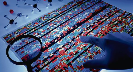
Un macroproyecto revela el mapa de los ‘interruptores’ del genoma

Fuente: http://elpais.com/elpais/2015/02/18/ciencia/1424249784_769402.html


El Proyecto Epigenoma Humano localiza las modificaciones del ADN que explican el desarrollo de la persona y sus grandes enfermedades.


El epigenoma humano es la síntesis del genoma y el entorno. 


Tras el genoma humano, llega el epigenoma humano. Lo que hace un gen no solo depende de su secuencia (gattacca…), sino de otras cosas que se pueden pegar sobre ella (de ahí epi, literalmente "encima de"), como los grupos más sencillos de la química orgánica (metilo, –CH3) y ciertas proteínas especializadas en empaquetar ADN (histonas). Estas modificaciones epigenéticas explican que, aunque todas las células de una persona tengan el mismo genoma, unas se conviertan en células de la piel, otras en neuronas, y así hasta los más de un centenar de tipos celulares especializados, o diferenciados, que constituyen el cuerpo.


Como las modificaciones epigenéticas dependen del entorno, también está cada vez más claro su papel esencial en todo tipo de respuestas al ambiente y enfermedades. Un macroproyecto revela hoy el mapa epigenómico humano, que localiza dónde están esas modificaciones (metilos, histonas) en todos los tipos celulares importantes del cuerpo humano, y también en 58 enfermedades (genéticamente) complejas. Como pasó con el proyecto Genoma Humano, la información se hará pública y gratuita para todo el mundo.


El macroestudio se presenta en varios artículos de Nature, Nature Communications, Nature Biotechnology, Nature Methods, Nature Neuroscience, Nature Immunology y Nature Protocols. Todo el material se puede consultar en una web habilitada para ello.


El principal coordinador del nuevo macroproyecto, Manolis Kellis, del Massachusetts Institute of Technology (MIT, Boston), amplía una metáfora clásica para explicar el concepto: “El proyecto genoma humano nos dio el libro de la vida que codifica a un ser humano. Todas nuestras células tienen una copia del mismo libro, pero cada una lee distintos capítulos, dobla la esquina de distintas páginas y subraya distintos párrafos y palabras. El epigenoma humano es esta colección de marcas situadas en el genoma de cada tipo celular, en la forma de modificaciones químicas del propio ADN, y en su empaquetamiento a gran escala”.


La numerología del proyecto se escapa en seguida al ámbito de las grandes cifras: más de 100 tejidos primarios y tipos celulares, 2.800 experimentos cada uno a escala del genoma entero, 150.000 millones de sondas o fragmentos de ADN que cubren el genoma 3.000 veces. Pero, como suele ocurrir, estas grandes cifras sirven sobre todo para marear al lector, o para impresionar al financiador. El proyecto epigenoma, probablemente, se puede entender mejor por sus objetivos a medio plazo, que se pueden expresar en términos más cualitativos.


Los investigadores de todo el mundo podrán a partir de ahora comparar la funcionalidad del genoma –qué genes están activos, activables o cerrados a conciencia— en cada tejido y tipo celular del cuerpo, consiguiendo un entendimiento profundo de lo que hace diferente a una célula del corazón de una del hígado, o de la piel, o del cerebro. Y también dispondrán de una referencia normal con que comparar las distintas células y tejidos de sus pacientes, revelando así cuáles son los elementos de control que causan su enfermedad en cualquier tejido u órgano.


Ya en esta fase inicial, los investigadores han asociado las variantes genéticas conocidas (variaciones en la propia secuencia del ADN: gattacca….) con su funcionalidad en tipos celulares específicos. Por ejemplo, se sabía que cierta variante genética se asocia con una alta o baja estatura, pero no a qué se debe ese efecto; ahora se ha podido ver que esa variación está activa (o tiene efectos sobre la actividad de ciertos genes) en las células madre, que así mantienen durante más o menos divisiones celulares su capacidad de proliferación.


Del mismo modo, los científicos ya pueden ver que las variantes genéticas asociadas a la diabetes de tipo 1, la artritis reumatoide y la esclerosis múltiple están activas en las células del sistema inmune. Esto es lógico, puesto que esas tres enfermedades pertenecen a la categoría autoinmune, es decir, que están causadas por un funcionamiento anómalo o excesivo de las defensas, que se dedican atacar a las células pancreáticas, articulares o nerviosas del propio paciente. Pero la lógica no sirve para encontrar tratamientos, y lo que tenemos ahora son datos duros que sí lo permitirán.


En la misma línea, las variantes genéticas asociadas con la presión arterial están activas en las células del corazón; las asociadas con un alto nivel de colesterol –y por tanto con una probabilidad incrementada de infarto— funcionan en el hígado, como las asociadas a los trastornos metabólicos. Todo esto tiene su lógica también, desde luego, pero de nuevo son los datos duros, y no la pura lógica, la que permitirá avanzar en el conocimiento de estas enfermedades y buscar tratamientos eficaces.


Un buen ejemplo de cómo el epigenoma puede ayudar a la medicina es el alzheimer. Puesto que se trata de una enfermedad neurodegenerativa –que va matando neuronas del cerebro progresivamente—, cabría esperar que las variantes genéticas que lo favorecen estuvieran activas en las neuronas, causando que estas células acumularan los depósitos de proteínas tóxicos que las van matando. Pero no es así. Esas variantes funcionan en las células del sistema inmune: en concreto en las células denominadas monolitos CD14. Esto cambia por completo la perspectiva para investigar tratamientos preventivos o paliativos del alzheimer.


En cualquier caso, y como también ocurrió con la presentación del genoma humano en 2002, el epigenoma que se presenta ahora es sobre todo un recurso para la investigación, que revelará sus beneficios en años y décadas por venir. El ser humano, en la salud y la enfermedad, es hoy un poco más computable que ayer a esta misma hora.


Researchers generate a reference map of the human epigenome

Fuente: http://medicalxpress.com/news/2015-02-human-epigenome.html


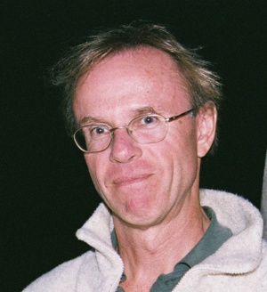
Manolis Kellis.


The sequencing of the human genome laid the foundation for the study of genetic variation and its links to a wide range of diseases. But the genome itself is only part of the story, as genes can be switched on and off by a range of chemical modifications, known as "epigenetic marks."


Now, a decade after the human genome was sequenced, the National Institutes of Health's Roadmap Epigenomics Consortium has created a similar map of the human epigenome.


Manolis Kellis, a professor of computer science and a member of MIT's Computer Science and Artificial Intelligence Laboratory and of the Broad Institute, led the effort to integrate and analyze the datasets produced by the project, which constitute the most comprehensive view of the human epigenome to date.


In a paper published in the journal Nature, Kellis and his colleagues report 111 reference human epigenomes and study their regulatory circuitry, in a bid to understand their role in human traits and diseases.


"The consortium set out to systematically characterize the human epigenomic landscape, across diverse tissues and cell types," Kellis says. "Given the enormity of the task, that meant bringing together multiple mapping centers and profiling a wide range of cell and tissue samples, to capture the diversity of the human epigenome." 


The researchers generated 2,805 genome-wide datasets, encompassing a total of 150 billion sequencing reads, corresponding to 3,174-fold coverage of the human genome. These captured modifications of both the DNA itself, and of the histone proteins around which DNA is wrapped to form a structure known as chromatin.


Kellis and his team then developed and applied machine-learning algorithms that could translate these datasets into a reference map in each of the 111 cell types and tissues. The algorithms distinguished different classes of epigenomic modifications and used them to annotate the genomic regions active in each sample, and in particular regulatory elements that control where and when different genes are expressed.


"Different combinations of epigenetic marks characterize different regions of the genome, reflecting the specific functions that they play in each cell," Kellis says. "By studying these combinations systematically, we can learn the language of the epigenome, and what it is telling us about both the activity and the function of each genomic region in each of the cell types."


The researchers distinguished 15 different epigenomic signatures, or chromatin states, reflecting active, repressed, poised, transcribed, and inactive regions of the genome in each cell type. About 5 percent of each reference epigenome showed signatures associated with a regulatory function.


"Chromatin states allowed us to summarize the complexity of diverse epigenomic marks into a small number of common patterns," Kellis says. "We could then interpret the biological functions of these patterns."


The researchers then studied how these chromatin states varied across different types of cells and tissues. This allowed them to group cell types with similar regulatory circuitry. They also grouped together regulatory regions that are active in the same types of cells. In this way they could begin to reveal the building blocks of regulatory circuits.


"Unlike the genome, which is mostly unchanged across cell types, the epigenome is extremely dynamic, reflecting the specialization of each cell type, such as neurons, heart, muscle, liver, skin, blood, or immune cells," Kellis says. "By studying which regions turn on and off in the same cell types, we can gain insights into gene regulation."


The researchers grouped 2 million predicted regulatory regions into 200 sets, or modules, which appeared to be acting in a coordinated manner across different types of cells. They found that 100 of these modules contained common sequence patterns, known as regulatory motifs, which may be responsible for their ability to work together in this way.


"Exploiting the predicted regulators and their motifs can help dissect the circuitry of different tissues and cells," Kellis says.


The researchers also compared these epigenomic signatures with groups of genetic variants that are associated with different human traits and diseases. This allowed them to produce a map of the tissue and cell types that are most relevant to each trait or disease.


"We found that genetic variants are found in regulatory regions known as enhancers, which are activated only in certain types of cell and tissue," Kellis says. "This suggests that many genetic variants affect the regulatory circuitry of the cell, possibly disrupting gene functions by altering tissue-specific gene expression levels."


The researchers found significant tissue-specific enhancer signatures for genetic variants associated with 58 different traits. These included height, in embryonic stem cells; multiple sclerosis, in immune cells; attention deficit disorder, in brain tissues; blood pressure, in heart tissues; fasting glucose, in pancreatic islets; cholesterol, in liver tissue; and Alzheimer's disease, in CD14 monocytes.


"This unbiased view allows researchers to focus on relevant cells and tissues that may have been otherwise overlooked when studying a particular disease," Kellis says. "The regulatory circuitry of a diverse range of cells can contribute to diseases that manifest in seemingly unexpected organs."


Using these circuits to understand the molecular basis of human disorders will take many years and the effort of many labs, Kellis says. "Our results provide an invaluable map, and a rich set of hypotheses, which can help guide these studies."


Wolf Reik, head of the epigenetics research program at the Babraham Institute in the U.K., who was not involved in the research, says the project is an exciting resource for the biomedical community worldwide. 


"Important epigenetic marks were mapped systematically in many human cell types and tissues," Reik says. "Integrative analysis of these epigenomes provides a global map toward understanding fundamental developmental and disease processes in humans."


More information: 


The research will be freely available to all, and the papers from Nature, along with papers from Nature Communications, Nature Biotechnology, Nature Methods, Nature Neuroscience, Nature Immunology and Nature Protocols, can be explored on the Epigenome Roadmap Site at www.nature.com/epigenomeroadmap.


Journal reference: Nature


domingo, 15 de febrero de 2015

Una carambola genética en una de sus células cura a una mujer enferma

Fuente: http://elpais.com/elpais/2015/02/05/ciencia/1423137442_823380.html


Un 'terremoto' natural en el ADN provoca por primera vez una curación espontánea.


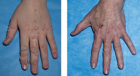
Mano de la mujer curada (derecha) y mano de su hija enferma.


“Lo más increíble de los milagros es que ocurren", decía un personaje del escritor británico G. K. Chesterton. Y, aparentemente, es cierto. Uno de ellos le ocurrió a una mujer de EE UU, denominada WHIM-09 por sus médicos para proteger su privacidad. Había nacido con una rarísima enfermedad congénita, con apenas 65 casos conocidos en el mundo, que destrozaba las defensas de su organismo y permitía que entraran en tromba virus y bacterias. Hasta los 38 años, una catarata de infecciones convirtió el hospital en su segunda casa. Su cuerpo además estaba lleno de verrugas, amenazando con degenerar en un cáncer. Pero un día, de repente, se curó.


Fue un presunto milagro, pero hoy, 20 años después de la supuesta intervención divina, un equipo de científicos explica la increíble carambola natural que en realidad curó a la señora. La mujer nació con una mutación en uno de sus 25.000 genes, el CXCR4, relacionado con las células madre que en el interior de los huesos forman la sangre, incluidas las defensas ante las infecciones. Era un cambio en solo una de las dos copias que los humanos tenemos de cada gen, pero esta pequeña tara provocaba su terrible enfermedad, conocida como síndrome WHIM.


Hace dos décadas, sin embargo, la paciente, por entonces con 38 años de infecciones a sus espaldas, sufrió otro extraño fenómeno: la cromotripsis. En cada célula del cuerpo humano, nuestros 25.000 genes se distribuyen en 23 paquetes duplicados o cromosomas. Cuando ocurre una cromotripsis, uno de estos cromosomas, espontáneamente, revienta en pedazos en una sola célula y sus cachitos se vuelven a recolocar como pueden. En muchos casos, esta catástrofe microscópica acaba con la muerte de la célula y pasa desapercibida. En otros, la célula se convierte en un monstruo, se multiplica y acaba provocando un cáncer.


En la mujer de EE UU, sin embargo, la explosión de un cromosoma en una de sus células salió de maravilla. En la hecatombe, su gen mutado desapareció junto a otros 163 genes, y el resto del cromosoma se volvió a unir de manera aleatoria. “La célula en la que ocurrió el fenómeno en nuestra paciente ya no era capaz de provocar el síndrome WHIM porque le faltaba el gen enfermo”, detalla el inmunólogo Philip M. Murphy, cuyo equipo describe el caso en la revista Cell. Es un proceso similar a tirar por los aires un plato de sopa de letras y que caiga al suelo formando la lista de ganadores de los premios Oscar de este año.


Murphy compara el fenómeno con el trencadís, los mosaicos de fragmentos de baldosas cerámicas utilizados por el arquitecto español Antoni Gaudí. "Es muy irónico que la paciente sea bordadora de colchas y edredones, una labor que puede considerarse una versión en tela del trencadís y una buena metáfora de la cromotripsis", apunta el investigador.


“Al cabo del tiempo, la célula con cromotripsis reemplazó a todas las células no saludables y formadoras de sangre que eran responsables del síndrome WHIM. Como resultado, los síntomas de la paciente desaparecieron y ha estado sana durante unos 20 años. La damos por curada”, añade Murphy, de los Institutos Nacionales de la Salud, en Bethesda (EE UU). Los investigadores se percataron de la causa del presunto milagro cuando la mujer, con 58 años, acudió a su centro con dos de sus hijas para someterse a un chequeo. Las chicas, de 21 y 23 años, presentaban los síntomas del síndrome. Los de la mujer habían desaparecido dos décadas antes.


“Esto es una terapia génica natural a partir de un fenómeno normalmente asociado con el cáncer, por casualidad total”, opina Juan Cruz Cigudosa, experto en aberraciones cromosómicas del Centro Nacional de Investigaciones Oncológicas (CNIO). Se calcula que hasta un 2% de todos los cánceres está asociado a la cromotripsis, a la explosión espontánea de un cromosoma. Esta es la primera vez que se documenta un caso en el que la catástrofe celular cura una enfermedad, según los autores.


El inmunólogo Philip M. Murphy.


“Ahora que sabemos que la cromotripsis puede curar una enfermedad, puede que los médicos que tienen pacientes con aparentes curaciones milagrosas quieran considerar este fenómeno como una posible explicación natural”, señala Murphy.


El bioquímico Óscar Fernández Capetillo, jefe del grupo de Inestabilidad Genómica del CNIO, recuerda que algunos investigadores han propuesto una infección vírica como explicación de la explosión cromosómica, pero él considera más plausible que se deba a un fallo en la división de una célula. "Lo interesante sería secuenciar el genoma de personas que se curan de cáncer espontáneamente y para las que no hace falta invocar al Espíritu Santo", apunta.


La terapia génica, la versión artificial de este evento natural, es un tratamiento innovador que ya se está probando con éxito contra algunas enfermedades, como la de los llamados niños burbuja, obligados a vivir aislados del mundo por una mutación en uno de sus genes relacionados con las defensas del organismo. El año pasado, investigadores del Centro de Oncología y Enfermedades de la Sangre Dana-Farber, en Boston (EEUU), anunciaron que habían extraído células madre productoras de sangre a ocho niños burbuja, les habían añadido una copia correcta del gen y se las habían vuelto a inyectar por millones. Los niños vivían casi con normalidad al cabo de entre uno y tres años después del tratamiento.


Para Murphy, el insólito caso de la mujer de EE UU puede servir para mejorar las terapias génicas e incluso los trasplantes de médula ósea para personas con leucemia. En su estudio, Murphy y sus colegas muestran, confirmándolo en ratones, que “tener una sola copia del gen CXCR4 es mejor que tener dos copias a la hora de que se injerten las células madre” en la médula ósea del interior de los huesos. Eso explicaría el éxito de la célula que sufrió la catástrofe cromosómica y acabó imponiéndose a todas las demás.


La aplicación potencial, por lo tanto, sería inactivar una copia del gen CXCR4 en cada célula madre antes de un trasplante a una persona con leucemia. Así, teóricamente, se facilitaría el anidamiento de las células del donante en la médula ósea del enfermo. Sin embargo, otros expertos son escépticos, como el farmacólogo Juan Bueren, experto en terapias innovadoras en el organismo público español CIEMAT.


“Yo no creo que se pueda concluir de este trabajo que las células madre con una copia inactivada del gen CXCR4 se injerten mejor, creo que proliferan más”, sostiene Bueren. El matiz no es baladí. El 90% de las células madre que producen sangre en un paciente sano están “dormidas, para protegerse y tener más tiempo para reparar fallos en su ADN”, recuerda el experto. Forzar su proliferación supondría “un estrés continuo” que podría generar efectos indeseados, como la anemia, afirma Bueren, que pide estudios a largo plazo para bendecir esta estrategia. Por el momento, el supuesto milagro solo beneficiará a la mujer conocida como WHIM-09.


sábado, 8 de noviembre de 2014

Scientists discover new step in a molecular pathway responsible for birth defects

Fuente: http://medicalxpress.com/news/2014-11-scientists-molecular-pathway-responsible-birth.html


Mary R. Loeken, Ph.D., Investigator in the Section on Islet Cell and Regenerative Biology at Joslin and Associate Professor of Medicine at Harvard Medical School. 


Mary R. Loeken, Ph.D., Investigator in the Section on Islet Cell and Regenerative Biology at Joslin Diabetes Center and Associate Professor of Medicine at Harvard Medical School, has discovered a molecular pathway responsible for neural tube defects in diabetic pregnancies. Her latest research findings in this pathway were published in the issue of Diabetes.


For 20 years, scientists have known of a gene involved in neural tube defects (such as spina bifida), but until now it was not known exactly what causes this gene to malfunctions during diabetic pregnancies.


A neural tube is the body's first step in assembling the spinal cord and the brain, which takes place within the first two to four weeks of gestation. Since the early 1990's, scientists have known that the gene Pax3 is required for closure of the neural tube.


Diabetic women who become pregnant run a greater than average risk that their baby, while the tube is folding shut, will develop anencephaly, a fatal disorder, or spina bifida, which can lead to motor impairment and other disabilities. For over two decades Dr. Loeken has focused her research on discovering how and why the malfunction of Pax3 can occur in diabetic women when they are pregnant.


In previous research, she found that Pax3 is sensitive to high levels of glucose. When exposed to hyperglycemic events, Pax3 doesn't turn on as much as it is supposed to. This is particularly dangerous as it occurs during the first month of pregnancy when a woman with diabetes may not be aware that she is pregnant and therefore may not take measures to have good control of her blood glucose.


In her paper this month in Diabetes, Dr. Loeken describes what prevents Pax3 from switching on. The key is an enzyme called DNA Methyltransferase (Dnmt), specifically subtype Dnmt 3b. DNA Methyltransferases are a family of enzymes responsible for altering the chemical building blocks of DNA, specifically adding a methyl group to cytosine (one of the four main bases in DNA's "rungs").


In normal development, Dnmt adds methyl groups to DNA around the Pax3 gene and then abruptly tapers off before the Pax3 gene comes on. Dr. Loeken discovered that glucose over-stimulates the DNA Methyltransferase 3b. The overactive enzyme keeps adding methyl groups to the cytosines near the Pax3 gene and prevents Pax3 from turning on so that it can close the neural tube.


Although it would appear that the way to prevent neural tube defects would be to stop Dnmt3b, Dr. Loeken cautions against such radical treatment. "There are actually drugs that can be used to inhibit DNA methylation; however we wouldn't use a drug to interfere with this pathway because this is a tightly regulated process."


According to Dr. Loeken, Dnmt3b isn't an entirely bad enzyme; there are many genes critical for development that need to be switched on or off by DNA methyltransferases. Dnmt3b is essential for embryonic survival. "However," says Dr. Loeken, "It does allow us to elucidate a biochemical pathway that we really didn't understand very well before."


A treatment that may come out of this research is better stem cell therapy. "We might be able to exploit these pathways to make more competent stem cells to repair the congenital malformations," says Dr. Loeken. "And that's not just limited to diabetes."


Neural tube defects happen in non-diabetic pregnancies as well, affecting about 1,500 births in the US every year and around 300,000 worldwide. Dr. Loeken's research has already caught the attention of other scientists working on neural tube defects. Together, they may illuminate causes and treatments for all babies who develop these malformations.


Until a treatment is developed, the best method for safeguarding against complications in diabetic pregnancies, Dr. Loeken points out, is to rigidly control a pregnant woman's blood glucose levels beginning before she is pregnant, and to plan their pregnancies.


"If the mother is under very tight glycemic control and avoids excursions above the level that will drive glucose into the cells, then all the events downstream, including the hypermethylation, should not occur."


More information: Wei, D & Loeken, M. Increased DNA methyltransferase 3b (dnmt3b)-mediated CpG island methylation stimulated by oxidative stress inhibits expression of a gene required for neural tube and neural crest development in diabetic pregnancy. Diabetes; vol 63, pp. 3512-22, October 2014


domingo, 12 de octubre de 2014

Special chromosomal structures control key genes

Fuente: http://www.sciencecodex.com/special_chromosomal_structures_control_key_genes-143237


Within almost every human cell is a nucleus six microns in diameter—about one 300th of a human hair's width—that is filled with roughly three meters of DNA. As the instructions for all cell processes, the DNA must be accessible to the cell's transcription machinery yet be compressed tightly enough to fit inside the nucleus. Scientists have long theorized that the way DNA is packaged affects gene expression. Whitehead Institute researchers present the first evidence that DNA scaffolding is responsible for enhancing and repressing gene expression.

"For the first time, we see that the structure of the chromosomes contributes to gene control," says Whitehead Member Richard Young, who is also a professor of biology at MIT. "In the past, there have been all kinds of ideas around how the structure might affect gene control, but now one has been experimentally tested and shown to be true."

DNA scaffolding has a hierarchy that ranges from beads-on-a-string, where a strand of DNA is wrapped around histone proteins to form a nucleosome, to topological associating domains akin to DNA balls containing multiple DNA loops interacting with various regulatory elements, to highly condensed chromosomes.

Working in mouse embryonic stem cells (ESCs), the Young lab examined the scaffolding landscape found within topologically associated domains. The researchers knew that proteins called Cohesin and CTCF are bound to the DNA around important ESC genes, but they did not know how these proteins affect the DNA's three-dimensional organization. Using a technique known as ChIA-PET, the researchers focused on how these proteins interact.

"By knowing which of the Cohesin/CTCF bound sites are coming together in physical proximity, we started to go from a linear view of the genome to sets of looping interactions, which led us to these domains, these super enhancer domains, where gene expression enhancement is contained within the loop," says Jill Dowen, a postdoctoral researcher in Young's lab.

Dowen and coauthors Zi Peng Fan, Denes Hnisz, and Gang Ren describe the structure of these loops in the latest online edition of the journal Cell.

Hnisz, also a postdoctoral researcher in Young's lab, likens the loops to "goody bags", with Cohesin and CTCF acting as the purse strings to create a DNA loop that cradles proteins enhancing or repressing gene expression. The enhancement or repression effects are confined to the genes within the goody bag. In the ESCs they studied, the scientists identified 197 Cohesin/CTCF-flanked loops that contain active genes and enhancers, and 349 loops that contain repressed genes.

Interestingly, when mouse neural precursor cells were examined for similar Cohesin/CTCF-bound loops, not only did the cells have such loops, but they were in similar locations as in the ESC genome. Of course, the repression or enhancement role of the loops differed between ESCs and the more differentiated cells. This indicates that the loops may remain constant during a cell's differentiation, although they may switch their contents, and therefore their effect on gene expression.

The researchers plan to study the loop structure and placement in other types of differentiated cells, but for now, they are excited about their new appreciation of DNA structure and its effects.

"I think we've filled in a major gap in the understanding of how gene expression is linked to the local organization of chromosomes," says Hnisz. "It will be exciting to explore whether defects in these mechanisms might even contribute to human disease."


Multiple neurodevelopmental disorders have a common molecular cause

Fuente: http://www.sciencecodex.com/multiple_neurodevelopmental_disorders_have_a_common_molecular_cause-143243


Neurodevelopmental disorders such as Down syndrome and autism-spectrum disorder can have profound, lifelong effects on learning and memory, but relatively little is known about the molecular pathways affected by these diseases. A study published by Cell Press in the American Journal of Human Genetics shows that neurodevelopmental disorders caused by distinct genetic mutations produce similar molecular effects in cells, suggesting that a one-size-fits-all therapeutic approach could be effective for conditions ranging from seizures to attention-deficit hyperactivity disorder.

"Neurodevelopmental disorders are rare, meaning trying to treat them is not efficient," says senior study author Carl Ernst of McGill University. "Once we fully define the major common pathways involved, targeting these pathways for treatment becomes a viable option that can affect the largest number of people."

A large fraction of neurodevelopmental disorders are associated with variation in specific genes, but the genetic factors responsible for these diseases are very complex. For example, whereas common variants in the same gene have been associated with two or more different disorders, mutations in many different genes can lead to similar diseases. As a result, it has not been clear whether genetic mutations that cause neurodevelopmental disorders affect distinct molecular pathways or converge on similar cellular functions.

To address this question, Ernst and his team used human fetal brain cells to study the molecular effects of reducing the activity of genes that are mutated in two distinct autism-spectrum disorders. Changes in transcription factor 4 (TCF4) cause 18q21 deletion syndrome, which is characterized by intellectual disability and psychiatric problems, and mutations in euchromatic histone methyltransferase 1 (EHMT1) cause similar symptoms in a disease known as 9q34 deletion syndrome.

Interfering with the activity of TCF4 or EHMT1 produced similar molecular effects in the cells. Strikingly, both of these genetic modifications resulted in molecular patterns that resemble those of cells that are differentiating, or converting from immature cells to more specialized cells. "Our study suggests that one fundamental cause of disease is that neural stem cells choose to become full brain cells too early," Ernst says. "This could affect how they incorporate into cellular networks, for example, leading to the clinical symptoms that we see in kids with these diseases."


Source: Cell Press

viernes, 1 de agosto de 2014

No extra mutations in modified stem cells, study finds

Fuente: http://www.salk.edu/news/pressrelease_details.php?press_id=2036


New results ease previous concerns that gene-editing techniques—used to develop therapies for genetic diseases—could add unwanted mutations to stem cells.


The ability to switch out one gene for another in a line of living stem cells has only crossed from science fiction to reality within this decade. As with any new technology, it brings with it both promise—the hope of fixing disease-causing genes in humans, for example—as well as questions and safety concerns. Now, Salk scientists have put one of those concerns to rest: using gene-editing techniques on stem cells doesn't increase the overall occurrence of mutations in the cells. The new results were published in the journal Cell Stem Cell.

"The ability to precisely modify the DNA of stem cells has greatly accelerated research on human diseases and cell therapy," says senior author Juan Carlos Izpisua Belmonte, professor in Salk's Gene Expression Laboratory. "To successfully translate this technology into the clinic, we first need to scrutinize the safety of these modified stem cells, such as their genome stability and mutational load."


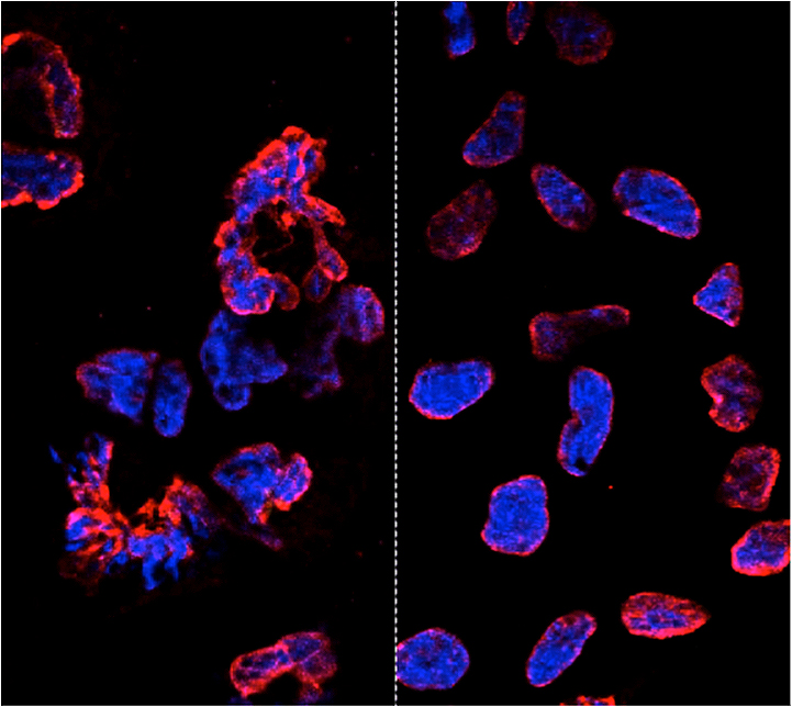
The new study shows that gene-editing technologies are specific to their targets and do not introduce harmful mutations, clearing the way for the development of safe therapies in the clinic. The left panel shows misshapen nuclear envelopes (red) from induced pluripotent stem cells derived from cells with Parkinson's disease (DNA in blue). The right panel shows similarly induced cells that have been gene-edited to restore the cells.

Click here for a high-resolution image.

Image: Courtesy of the Salk Institute for Biological Studies


When scientists want to change the sequence of a stretch of DNA inside cells—either for research purposes or to fix a genetic mutation for therapeutic purposes—they have their choice of two methods. They can use an engineered virus to deliver the new gene to a cell; the cell then integrates the new DNA sequence in place of the old one. Or scientists can use what's known as custom targeted nucleases, such as TALEN proteins, which cut DNA at any desired location. Researchers can use the proteins to cut a gene they want to replace, then add a new gene to the mix. The cell's natural repair mechanisms will paste the new gene in place.


Previously, Belmonte's lab had pioneered the use of modified viruses, called helper-dependent adenoviral vectors (HDAdVs) to correct the gene mutation that causes sickle cell disease, one of the most severe blood diseases in the world. He and his collaborators used HDAdVs to replace the mutated gene in a line of stem cells with a mutant-free version, creating stem cells that could theoretically be infused into patients' bone marrow so that their bodies create healthy blood cells.

Before such technologies are applied to humans, though, researchers like Belmonte wanted to know whether there were risks of editing the genes in stem cells. Even though both common gene-editing techniques have been shown to be accurate at altering the right stretch of DNA, scientists worried that the process could make the cells more unstable and prone to mutations in unrelated genes—such as those that could cause cancer.

"As cells are being reprogrammed into stem cells, they tend to accumulate many mutations," says Mo Li, a postdoctoral fellow in Belmonte's lab and an author of the new paper. "So people naturally worry that any process you perform with these cells in vitro—including gene editing—might generate even more mutations."


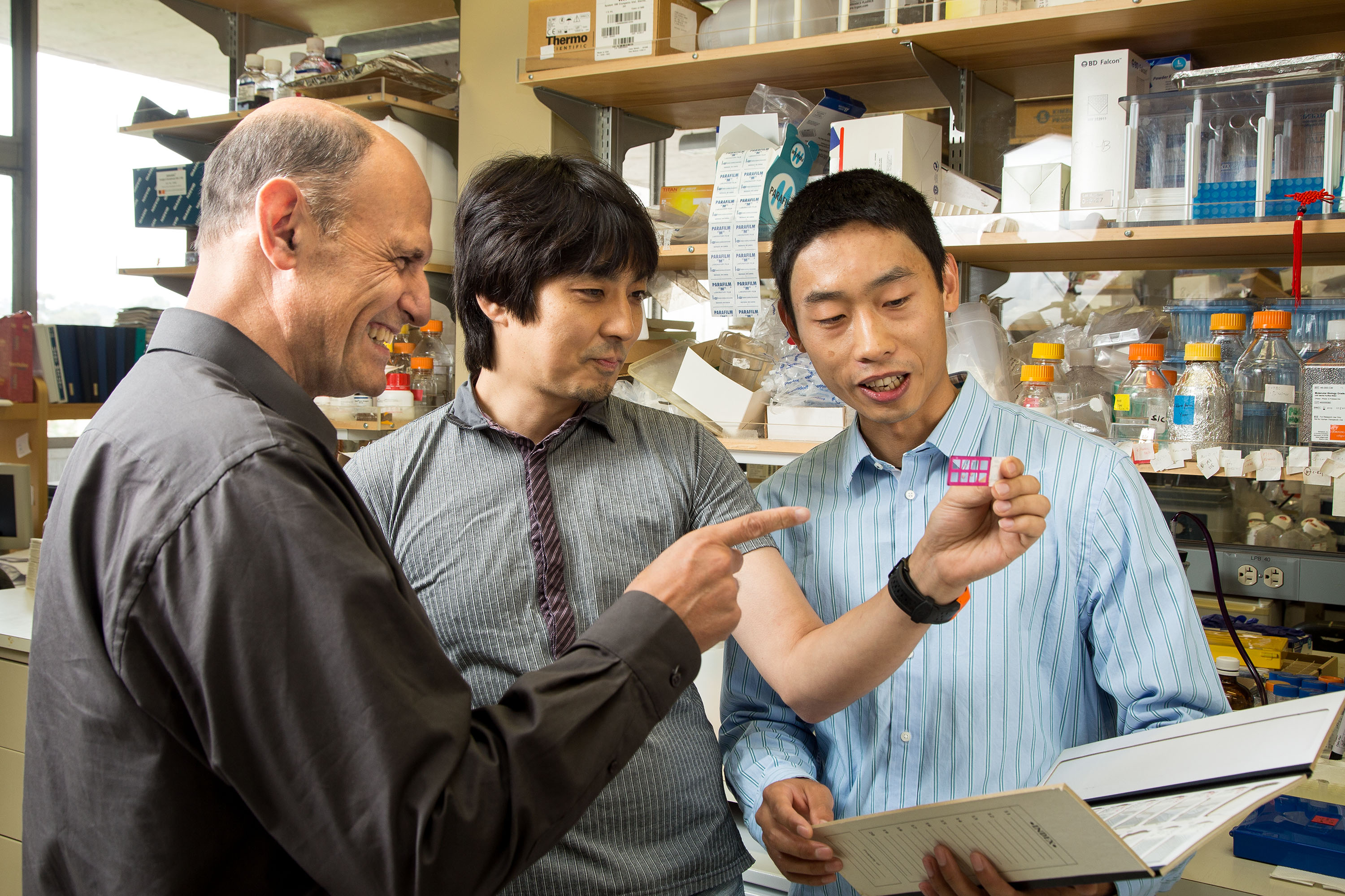
From left: Juan Carlos Izpisua Belmonte, Keiichiro Suzuki and Mo Li of the Gene Expression Laboratory

Click here for a high-resolution image.

Image: Courtesy of the Salk Institute for Biological Studies


To find out whether this was the case, Belmonte's group, in collaboration with BGI and the Institute of Biophysics, Chinese Academy of Sciences in China, turned to a line of stem cells containing the mutated gene that causes sickle cell disease. They edited the genes of some cells using one of two HDAdV designs, edited others using one of two TALEN proteins, and kept the rest of the cells in culture without editing them. Then, they fully sequenced the entire genome of each cell from the four edits and control experiment.

While all of the cells gained a low level of random gene mutations during the experiments, the cells that had undergone gene-editing—whether through HDAdV—or TALEN-based approaches—had no more mutations than the cells kept in culture.

"We were pleasantly surprised by the results," Keiichiro Suzuki, a postdoctoral fellow in Belmonte's lab and an author of the study, says. "People have found thousands of mutations introduced during iPSC reprogramming. We found less than a hundred single nucleotide variants in all cases."

The finding, Li adds, doesn't necessarily mean that there are no inherent risks to using stem cells with edited genes, but that the editing process doesn't make the stem cells any less safe.

"We concluded that the risk of mutation isn't inherently connected to gene editing," he says. "These cells present the same risks as using any other cells manipulated for cell or gene therapy." He adds that two other papers published in the same issue support their results (one by Johns Hopkins University and one from Harvard University and collaborators).

The Belmonte group is planning more studies to address whether gene-repair in other cell types, using other approaches, or targeting other genes could be more or less likely to cause unwanted mutations. For now, they hope their findings encourage those in the field to keep pursuing gene-editing techniques as a potential way to treat genetic diseases in the future.

Other researchers on the study were Jing Qu, April Goebl, Emi Aizawa, Rupa Devi Soligalla, Jessica Kim, Na Young Kim, Hsin-Kai Liao, Chris Benner, and Concepcion Rodriguez Esteban of the Salk Institute for Biological Studies; Chang Yu, Xiaotian Yao, Senwei Tang, Fan Zhang, Feng Chen, Yabin Jin, and Yingrui Li of BGI; and Jing Qu,Tingting Yuan, Ruotong Ren, Xiuling Xu, and Guang-Hui Liu of the Institute of Biophysics, Chinese Academy of Sciences.


About the Salk Institute for Biological Studies:

The Salk Institute for Biological Studies is one of the world's preeminent basic research institutions, where internationally renowned faculty probes fundamental life science questions in a unique, collaborative and creative environment. Focused both on discovery and on mentoring future generations of researchers, Salk scientists make groundbreaking contributions to our understanding of cancer, aging, Alzheimer's, diabetes and infectious diseases by studying neuroscience, genetics, cell and plant biology, and related disciplines.

Faculty achievements have been recognized with numerous honors, including Nobel Prizes and memberships in the National Academy of Sciences. Founded in 1960 by polio vaccine pioneer Jonas Salk, MD, the Institute is an independent nonprofit organization and architectural landmark.

domingo, 27 de julio de 2014

Gene discovered that activates stem cells for organ regeneration in planarians

Fuente: http://esciencenews.com/articles/2014/07/04/gene.discovered.activates.stem.cells.organ.regeneration.planarians


Researchers announced the discovery of a gene zic-1 that enables stem cells to regrow a head after decapitation in flatworm planarians. Professor Christian Petersen and Ph.D. student Constanza Vásquez-Doorman of Northwestern University discovered zic-1 by investigating planarians, an animal that uses pluripotent stem cells to regrow any missing tissue lost from injury. The study, entitled "zic-1 Expression in Planarian Neoblasts after Injury Controls Anterior Pole Regeneration," was reported in PLOS Genetics.

Many species across the animal kingdom have the ability to regenerate, but the mechanisms that connect injuries to stem cell activation and the production of new tissues are not fully understood. Developmental biologists have established that in the early embryo most animals use "tissue organizers" that secrete proteins to allow cell-cell communication for the formation of organs. But it was not clear how such tissue organizers could be produced from scratch to allow adult regeneration in animals like planarians.

A clue to the solution came from Petersen's previous research that identified the secreted protein Notum as a component of a tissue organizer needed for head regeneration in planarians. Notum drives head regeneration by inhibiting Wnt signaling, a broadly used molecular pathway used in cell-cell communication. Vásquez-Doorman and Petersen found that expression of the Notum protein and head regeneration required the zic-1 gene, which encodes a DNA-binding protein activated in stem cells early after decapitation.

Human stem cells could ultimately be used to build or repair complex tissues, but most ongoing research on these cells has focused on their ability to create individual cell types in a dish. Petersen's study identifies a potentially ancient connection between Wnt signaling and zic-1 in the use of stem cells for coordinating regenerative growth and suggests that human stem cells might someday be used to create tissue organizers for enhancement of injury repair.


Source: PLOS