Boston researchers have identified a way to enhance regrowth of human corneal tissue to restore vision, using a molecule known as ABCB5 that acts as a marker for hard-to-find limbal stem cells. This work, a collaboration between the Massachusetts Eye and Ear/Schepens Eye Research Institute (Mass. Eye and Ear), Boston Children's Hospital, Brigham and Women's Hospital and the VA Boston Healthcare System, provides promise to burn victims, victims of chemical injury and others with damaging eye diseases. The research, published in Nature, is also one of the first known examples of constructing a tissue from an adult-derived human stem cell.
Limbal stem cells reside in the eye's basal limbal epithelium, or limbus, and help maintain and regenerate corneal tissue. Their loss due to injury or disease is one of the leading causes of blindness. In the past, tissue or cell transplants have been used to help the cornea regenerate, but it was unknown whether there were actual limbal stem cells in the grafts, or how many, and the outcomes were not consistent.
In this study, researchers were able to use antibodies detecting ABCB5 to zero in on the stem cells in tissue from deceased human donors and use them to regrow anatomically correct, fully functional human corneas in mice.
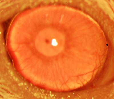
This is a restored functional cornea following transplantation of human ABCB5-positive limbal stem cells to limbal stem cell-deficient mice. Transplants consisting of human ABCB5-positive limbal stem cells resulted in restoration and long-term maintenance of a normal clear cornea, whereas control mice that received either no cells or ABCB5-negative cells failed to restore the cornea.
(Photo Credit: Kira Lathrop, Bruce Ksander, Markus Frank, and Natasha Frank.)
"Limbal stem cells are very rare, and successful transplants are dependent on these rare cells," says Bruce Ksander, Ph.D., of Mass. Eye and Ear, co-lead author on the study with post-doctoral fellow Paraskevi Kolovou, M.D. "This finding will now make it much easier to restore the corneal surface. It's a very good example of basic research moving quickly to a translational application."
ABCB5 was originally discovered in the lab of Markus Frank, M.D., of Boston Children's Hospital, and Natasha Frank, M.D., of the VA Boston Healthcare System and Brigham and Women's Hospital, co-senior investigators on the study, as being produced in tissue precursor cells in human skin and intestine. In the new work, using a mouse model developed by the Frank lab, they found that ABCB5 also occurs in limbal stem cells and is required for their maintenance and survival, and for corneal development and repair. Mice lacking a functional ABCB5 gene lost their populations of limbal stem cells, and their corneas healed poorly after injury.
"ABCB5 allows limbal stem cells to survive, protecting them from apoptosis [programmed cell death]," says Markus Frank. "The mouse model allowed us for the first time to understand the role of ABCB5 in normal development, and should be very important to the stem cell field in general." according to Natasha Frank.
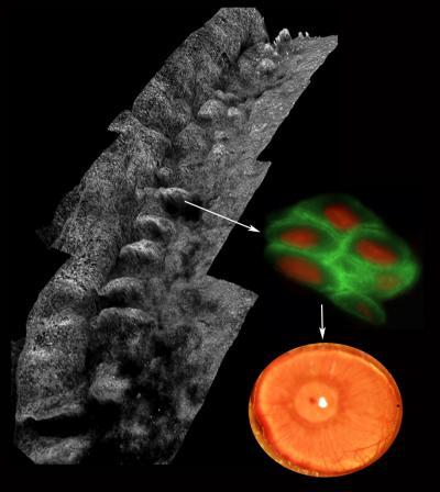
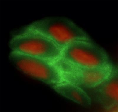
Composite image depicting the palisades of Vogt within the human limbus (left), ABCB5-positive limbal stem cells isolated from the palisades (right; ABCB5 -- green, nucleus -- red) and a restored functional cornea following transplantation of human ABCB5-positive limbal stem cells to limbal stem cell-deficient mice (bottom right).
(Photo Credit: Kira Lathrop, Bruce Ksander, Markus Frank, and Natasha Frank.)
Markus Frank is working with biopharmaceutical industry to develop a clinical-grade ABCB5 antibody that would meet U.S. regulatory approvals. "A single lab cannot do a study like this," says Natasha Frank, also affiliated with the Harvard Stem Cell Institute. "It integrates genetics, knockout mice, antibodies, transplantation—a lot of technical expertise that we were lucky came together in a very nice way."
This is an image depicting the palisades of Vogt within the human limbus. The human limbal architecture shows the palisades of Vogt in whole mounted tissue labeled with collagen VII and imaged with a laser scanning confocal microscope. Image is a stitched Z stack series.
(Photo Credit: Kira Lathrop, Bruce Ksander, Markus Frank, and Natasha Frank.)
No hay comentarios:
Publicar un comentario