jueves, 2 de octubre de 2014

Hallan células madre en el ojo que pueden reprogramarse para ser fotosensibles

Fuente: http://www.abc.es/salud/noticias/20141001/abci-celulas-madre-ceguera-terapia-201410011750.html


Ubicadas en el 'limbo corneal' están presentes incluso en personas de edad avanzada y podrían usarse para recuperar la visión.


La solución a muchas enfermedades relacionadas con la pérdida de visión podría estar en nuestros propios ojos. Científicos de la Universidad de Southampton (Gran Bretaña) han descubierto que una región en la superficie frontal del ojo alberga células madre especiales que podrían tratar afecciones oculares que provocan ceguera. La parte del ojo en cuestión es el ‘limbo corneal’ y es una especie de hueco estrecho que se extiende entre la córnea y la esclerótica transparente.

La investigación, publicada en «PLoS ONE», demuestra que las células madre pueden cultivarse in vitro desde el limbo corneal. Así, bajo las condiciones de cultivo adecuadas, estas células podrían ser programadas para comportarse como células fotorreceptoras, las células necesarias para poder ver la luz.


«Estas células son de fácil acceso y tienen sorprendente plasticidad, lo que las convierte en un recurso celular atractivo para futuras terapias», asegura Andrew Lotery, coordinador del trabajo. En su opinión, «esto ayudaría a evitar complicaciones como el rechazo o la contaminación debido a que las células son del propio paciente».

Otro dato importante de este trabajo es que estas células madre existen también en los ojos humanos de personas de edad avanzada y pueden cultivarse incluso desde el limbo corneal de una persona de 97 años de edad. Por lo tanto, «este descubrimiento abre la posibilidad de nuevos tratamientos para las personas mayores».

La pérdida de células fotorreceptoras provoca ceguera irreversible y los investigadores esperan que este descubrimiento podría conducir a nuevos tratamientos para patologías como la degeneración macular relacionada con la edad, la principal causa de ceguera en el mundo desarrollado.

Experts from CNIO discover shining cells responsible for developing tumors

Fuente: http://www.eurekalert.org/pub_releases/2014-09/cndi-efc092914.php


This finding will help to track the origin of chemical resistance and to launch personalized medicine by means of developing novel therapies against these cancer stem cells.


Researchers Bruno Sainz and Irene Miranda show the image of a tumor stem cell containing green-colored fluorescent vesicles.


Tumours are mosaics of cells that are morphologically and molecularly very different. In this cellular heterogeneity, it is calculated that only 1-2% of the tumour mass is made up of cancer stem cells, which over the past years have been suggested to be responsible for the origin of cancer and for the resistance to conventional chemical therapies. This small percentage of cancer stem cells in a solid tumour makes it difficult to isolate and analyse them, as well as to study the origin of drug resistance.


Researchers from the Spanish National Cancer Research Centre (CNIO) have discovered and characterised a new specific marker for cancer stem cells: riboflavin, or vitamin B2, a pigment that emits green fluorescence as a result of its accumulation inside intracellular vesicles. This light emission property, acts to track, isolate, and later purify it, without the need for antibodies or other more costly and complex techniques.


Results from the research, headed by scientists Irene Miranda, Bruno Sainz and Christopher Heeschen, are published in the journal Nature Methods.


"The discovery of this new marker is a breakthrough, as it can select for tumour stem cells, which are the most invasive and chemical-resistant cancer cells. Autofluorescence allows these cells to be tracked in an easy, simple and inexpensive way, as well as to study the origin of the tumours' chemical resistance," states Irene Miranda, the article's first author.


"Normally, we only see the leaves of the tree represented by tumours, but we cannot make out the roots [the cancer stem cells], which are the true responsible parties for the progression and growth of tumours," illustrates Miranda.


The discovery −which was carried out in several types of tumours, including samples from patients with pancreas, liver, colon and lung cancer− raises a question: why do tumour stem cells accumulate vitamin B2?


In the article, researchers show that this is due to an increase in ABCG2, the protein responsible for the transport of vitamin B2 into intracellular vesicles, conferring luminosity to the cells. The factors behind this phenomenon are yet to be determined.


Despite unknowns regarding its nature, autofluorescence could help to launch future approaches in personalised medicine and to develop more effective anticancer treatments. "We will now be able to isolate autofluorescent cells from a biopsy and to test their sensitivity in a panel of experimental or marketed drugs," affirm Sainz and Heeschen. "In this way, we want to accelerate the identification of new drugs or their use in combination, in order to specifically destroy the patient´s cancer stem cells," says Sainz.


###


The study was funded by the European Union, the National Institute of Health Carlos III, the Spanish Ministry of Economy and Competitiveness and the "la Caixa" Foundation.


Reference article: Intracellular autofluorescence: a biomarker for epithelial cancer stem cells. Irene Miranda-Lorenzo, Jorge Dorado, Enza Lonardo, Sonia Alcala, Alicia G Serrano, Jenifer Clausell-Tormos, Michele Cioffi, Diego Megias, Sladjana Zagorac, Anamaria Balic, Manuel Hidalgo, Mert Erkan, Joerg Kleeff, Aldo Scarpa, Bruno Sainz, Jr & Christopher Heeschen. Nature Methods (2014). doi: 10.1038/nmeth.3112

Disease decoded: Gene mutation may lead to development of new cancer drugs

Fuente: http://www.eurekalert.org/pub_releases/2014-09/uom-ddg093014.php


The discovery of a gene mutation that causes a rare premature aging disease could lead to the development of drugs that block the rapid, unstoppable cell division that makes cancer so deadly.

Scientists at the University of Michigan and the U-M Health System recently discovered a protein mutation that causes the devastating disease dyskeratosis congenita, in which precious hematopoietic stem cells can't regenerate and make new blood. People with DC age prematurely and are prone to cancer and bone marrow failure.

But the study findings reach far beyond the roughly one in 1 million known DC patients, and could ultimately lead to developing new drugs that prevent cancer from spreading, said Jayakrishnan Nandakumar, assistant professor in the U-M Department of Molecular, Cellular, and Developmental Biology.

The DC-causing mutation occurs in a protein called TPP1. The mutation inhibits TPP1's ability to bind the enzyme telomerase to the ends of chromosomes, which ultimately results in reduced hematopoietic stem cell division. While telomerase is underproduced in DC patients, the opposite is true for cells in cancer patients.

"Telomerase overproduction in cancer cells helps them divide uncontrollably, which is a hallmark of all cancers," Nandakumar said. "Inhibiting telomerase will be an effective way to kill cancer cells."

The findings could lead to the development of gene therapies to repair the mutation and start cell division in DC patients, or drugs to inhibit telomerase and cell division in cancer patients. Both would amount to huge treatment breakthroughs for DC and cancer patients, Nandakumar said.

Nandakumar said that a major step moving forward is to culture DC patient-derived cells and try to repair the TPP1 mutation to see if telomerase function can be restored. Ultimately, the U-M scientist hopes that fixing the TPP1 mutation repairs telomerase function and fuels cell division in the stem cells of DC patients.

"It's conceivable that with the recent advancement in human genome-editing technology, we could, in the not-so-distant future, repair the mutation in hematopoietic stem cells in the bone marrow of DC patients," Nandakumar said.

The findings also reinforce how one tiny change in an amino acid chain can cause devastating health consequences.

"It was surprising to us that just deleting one single amino acid in a protein chain that is 544 amino acids long can result in such a severe disease," Nandakumar said.


###


First author Hande Kocak conducted the research in Nandakumar's lab in the Department of Molecular, Cellular, and Developmental Biology. She is in the Department of Human Genetics at the U-M Medical School. Co-author Dr. Catherine Keegan is Kocak's mentor and has appointments in the departments of Human Genetics and Pediatrics.

The study, "Hoyeraal-Hreidarsson syndrome caused by a germline mutation in the TEL patch of the telomere protein TPP1," appears in the journal Genes and Development.


Jayakrishnan Nandakumar: http://bit.ly/1nFwdMf 

Molecular, Cellular, and Developmental Biology: http://www.lsa.umich.edu/mcdb

Researchers working to regenerate heart and nerve cells

Fuente: http://medicalxpress.com/news/2014-09-regenerate-heart-nerve-cells.html


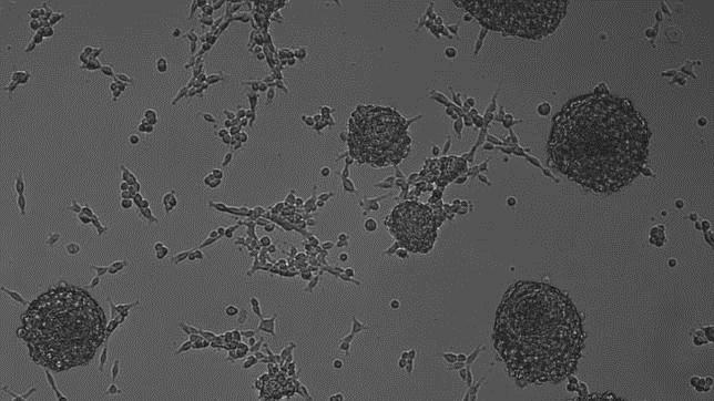
Under a microscope inside a lab at UT Southwestern Medical Center, a dish of cells pulsates with the rhythm of a human heart.


Days ago, these building blocks of heart muscle had a different identity altogether: They were scar-forming cells that proliferate in the wake of a heart attack and weaken the organ's ability to pump blood.


Researchers at UT Southwestern and other institutions may have found a way to reverse the damage that patients sustain from heart attacks, traumatic brain injuries and other conditions.


They accomplish this by converting a less desirable type of cell, such as a scar-forming cell, into a highly desirable one, such as a nerve cell, using a process known as direct reprogramming.


Most recent efforts to convert one cell type into another have relied on stem cells - immature versions of adult cells. But stem cells can be difficult to manipulate and have caused controversy because they are sometimes derived from embryos donated by fertility clinics.


Many scientists say direct reprogramming is a promising new approach to regenerative medicine - a field dedicated to curing disease by helping the body repair and regrow its own tissues.


In recent years, scientists have used the method to grow new brain cells, liver cells, pancreatic tissue and parts of the inner ear responsible for hearing loss.


Researchers hope to one day develop drugs that will enable patients' bodies to repair their own damaged organs, much the way a salamander can regrow its own heart and a python can double the size of its liver.


At UT Southwestern's new Hamon Center for Regenerative Science and Medicine, researchers are focused on cultivating heart and nerve cells.


The center, led by biologist Eric Olson, opened in May with a $10 million gift from the Hamon Charitable Foundation. Its areas of research include stem cells as well as direct reprogramming.


Olson hopes the center will help UT Southwestern attract top college graduates from around the U.S.


"Regenerative biology is one of the most popular majors," he says. "The center will give us a new tool to recruit some of the best students to Dallas."


Olson's career-long interest in how a single cell develops into the trillions that make up the human body led to his lab's current work.


"I wanted to understand how specialized cells were formed and how large sets of genes turned on and off during development," he said.


After pondering what cell type to focus on, Olson settled on muscle cells, because muscles make up 40 percent of our body mass and turn on thousands of genes as they develop.


The idea was that a deep understanding of one particular system could be applied more widely. It could also help scientists gain insight into how diseases develop when errors creep into the process.


Over the years, Olson's lab has uncovered several of the major genes that control muscle development.


Recent work from his lab has led to a promising approach to treating Duchenne muscular dystrophy, a congenital disease that causes muscles to progressively weaken and degenerate.


In the center's inaugural publication this month in the journal Science, Olson and his colleagues outlined a way to "edit" the gene abnormalities responsible for the disorder in a mouse and rectify the body's ability to grow healthy muscle.


Olson's lab extended its work into heart muscle cells in the early 1990s. At the time, Deepak Srivastava, then a postdoctoral researcher, was working in the lab.


"I was trained as a pediatric cardiologist and was interested in how the heart forms in the embryo and how that goes awry in human disease," says Srivastava, who now directs cardiac and stem cell research at the Gladstone Institutes and is a professor at the University of California San Francisco. The two went on to discover many of the genes that control heart development.


Using some of that research, Srivastava pioneered cardiac reprogramming several years later.


"We took the years of knowledge that our lab and other labs had developed about how nature normally makes a heart in the embryo and essentially redeployed those same methods in the adult heart," he says.


In a 2010 paper in the journal Cell, Srivastava showed that scar-forming cells known as fibroblasts could be converted into beating heart cells by adding just three ingredients. Those ingredients, proteins known as transcription factors, are master regulators that flip genes on and off. The genes involved are the same ones that direct heart formation in the womb.


In 2012, Srivastava and Olson, writing separate papers in the same issue of the journal Nature, showed that the process could be performed successfully in living mice.


Each group used a different combination of transcription factors to reprogram the cells, but both found that the technique improved heart function in the animals following a heart attack.


For the last two decades, stem cells have been virtually synonymous with regenerative medicine.


Scientists use them in many different ways. In one approach, researchers take a sample of skin, fat or bone marrow from a patient, convert the mature cells into their previous immature states, then reprogram them to form a new type of tissue.


In a second approach, scientists can extract stem cells from embryos and then convert them.


In clinical trials, researchers have also used a third approach: They have taken so-called adult stem cells from bone marrow and injected them into the heart, hoping the stem cells take on the properties of muscle cells.


Adult stem cells have had mixed results. "The reality is this has shown limited benefits after a decade of clinical trials," says Olson.


So far, researchers have tried direct reprogramming in mice and in human cells in a lab dish.


Chun-Li Zhang, a colleague of Olson's at the Hamon Institute, is using direct reprogramming to rebuild nerve cells, or neurons. He starts with glial cells, which reproduce in the brain and spinal cord following an injury and can form scars.


"Basically, these are the fibroblasts of the brain," he says.


In a series of papers published over the last year, Zhang and his colleagues reported generating new neurons from glial cells in mice following a traumatic brain or spinal cord injury. The lab is now investigating how much the new cells can contribute to the animals' recovery.


Most scientists who work on direct reprogramming are continuing their research on stem cells in parallel. "They each have advantages and disadvantages," says Srivastava.


George Daley, director of stem cell transplantation at Children's Hospital Boston and a past president of the International Society of Stem Cell Research, compared the effectiveness of the two methods in a paper published last month in the journal Cell.


Neurons and heart cells created using direct reprogramming "aren't as close to normal tissue as those generated from stem cells," he said.


Another advantage of using stem cells is that the process creates a lot more cells to work with than direct reprogramming.


"Right now, if you take a dish of fibroblasts in a lab and reprogram that dish, you can get maybe 10 percent of those cells to turn into heart muscle cells, and we'd like to improve that efficiency," says Olson.


Yet direct reprogramming, says Zhang, is more akin to true regeneration. "We design a way to help the tissue repair or regenerate itself," he says. Stem cells, by contrast, involve growing cells in a dish and then injecting them into patients.


The ultimate hope with direct reprogramming is that it will lead to a drug that will stimulate patients' bodies to heal themselves.


With both approaches, Food and Drug Administration-approved treatments are still at least several years away.


Srivastava is testing his direct reprogramming technique in pigs, whose hearts are closer in size to those of humans. The goal is to see if he can generate enough muscle to dramatically improve the heart's pumping ability.


If all goes well, he hopes to approach the FDA with a plan for clinical trials within two to three years.


"It's not inconceivable that we could know if this works in humans within the next five years," he says.


Journal reference: Cell Science


Multiple sclerosis patients successfully replace damaged cells with new healthy stem cells; first treatment with placenta

Fuente: http://www.medicaldaily.com/multiple-sclerosis-patients-successfully-replace-damaged-cells-new-healthy-stem-cells-305420


Multiple sclerosis is a chronic, life-altering incurable disease, but an unprecedented treatment may have opened doors up for a successful treatment using cells from human placenta tissues. Researchers from Mount Sinai designed the treatment and have found patients were able to handle the treatment, and published their results in the journal Multiple Sclerosis and Related Disorders.


Multiple sclerosis patients show improvement when damaged cells are replaced with placenta.


"This is the first time placenta-derived cells have been tested as a possible therapy for multiple sclerosis," the study’s lead author Fred Lublin, Director of the Corinne Goldsmith Dickinson Center for Multiple Sclerosis, said in a press release. "The next step will be to study larger numbers of MS patients to assess efficacy of the cells, but we could be looking at a new frontier in treatment for the disease."

When a patient is diagnosed with MS, an autoimmune disease that causes the body’s immune system to attack itself and disrupt the flow of information between the brain and the rest of the body, according to the National Multiple Sclerosis Society. More than 2.3 million people are affected by MS and while symptoms are unpredictable and can vary greatly in severity from one person to another, it always worsens overtime. The damaged nerve cells were repaired and tolerated by the MS person’s body successfully with PDA-001, a group of cultured placenta cells that resemble the connective tissue found in bone marrow. A placenta is an organ that encases the fetus inside a pregnant woman's womb, rich in oxygen and nutritents. By using cells cultured from placentas, researchers were able to extract significantly more of these cell-repairing cells from one donor, so they could supply many patients at a time.

Researchers examined the treatment in 16 patients with MS, some had relapsing-remitting multiple sclerosis (RRMS) and others had the more evolved version of the chronic and more debilitating condition called secondary progressive multiple sclerosis (SPMS). Patients were between the ages of 18 and 65 and one group was given a high dose of PDA-001, another were given a low dosage, and a third group was given a placebo. While there is always a risk for MS to worsen once the immune system is experimented with in cell transplants, over the six-month treatment the majority of the patients who were treated with PDA-001 had stabilized or showed improvement.

"We're hoping to learn more about how placental stromal cells contribute to myelin repair," Lublin said. "We suspect they either convert to a myelin making cell, or they enhance the environment of the area where the damage is to allow for natural repair. Our long-term goal is to develop strategies to facilitate repair of the damaged nervous system."


Source: Lublin F. Multiple Sclerosis and Related Disorders. 2014.

Células de sangre de pacientes infartados pueden regenerar el tejido dañado del corazón

Fuente: http://www.rtve.es/noticias/20140929/demuestran-celulas-sangre-paciente-infarto-pueden-restaurar-corazon/1019461.shtml


Las propias células de la sangre de pacientes que han sufrido un infarto de miocardio son capaces de restaurar el tejido dañado del corazón, según las conclusiones de un estudio realizado por investigadores de las Universidades de Granada, Jaén, Málaga, Miami y del Hospital Universitario Virgen de la Victoria de Málaga.

En concreto, se trata de las denominadas células progenitoras endoteliales (EPC), aquellas que están destinadas a convertirse en vasos sanguíneos, pero que los científicos han conseguido que se diferencien hacia células de músculo cardiaco (cardiomiocitos).

Esta posibilidad de cambiar su función se debe a que se encuentran en una fase inicial en la que las células aún no están maduras y, por tanto, tienen plasticidad, es decir, se pueden reconducir para se conviertan en tejido cardiaco.


“Hasta ahora se sabía el papel beneficioso de estas EPC en patologías donde se reduce el diámetro de los vasos sanguíneos como la cardiopatía isquémica y la enfermedad arterial periférica. Sin embargo, poco se sabía sobre su potencial para restaurar el tejido dañado del corazón”, ha explicado el responsable del proyecto en la Universidad de Granada, Juan Antonio Marchal.

Para esclarecer su papel, los científicos analizaron in vitro la capacidad de convertirse en células cardiacas de muestras de EPC aisladas a partir de sangre de pacientes con infarto agudo de miocardio.

Las compararon con EPC obtenidas a partir de sangre de cordón umbilical, una fuente donde ya se conoce que existen células madre capaces de regenerar tejido cardiaco.

“Es la primera vez que se desarrolla esta comparación. Nuestros datos indican que células progenitoras endoteliales (EPCs) obtenidas a partir de ambos orígenes, sangre de pacientes y cordón umbilical, tienen plasticidad y funciones similares y sugieren una eficacia terapéutica potencial en la terapia celular cardiaca”, ha aseverado el investigador.


Esta terapia pasaría por inyectar estas células en la zona de la lesión. “Tras un infarto, se produce un efecto llamada de estas EPC desde la médula osea, donde se encuentran normalmente, hasta el corazón, para reparar la lesión: es el efecto homing. Sin embargo, este mecanismo no logra trasladar suficientes células como para regenerar por completo el tejido dañado”, explica.


Por eso, la futura terapia que proponen los expertos ayudaría a los pacientes con ese proceso de regeneración. Además, al tratarse de células propias se evitan posibles rechazos.

“La inyección de células ya se hace, pero nosotros hemos demostrado la capacidad de estas células propias del paciente infartado, de ahí que no se produzcan efectos secundarios”, aclara.

“No obstante, es necesario desarrollar protocolos de expansión en el laboratorio de estas células del propio paciente para que podamos obtener un número adecuado que tuviera el efecto beneficioso una vez introducidas en los pacientes”, precisa.


Para llegar a sus conclusiones, publicadas en la revista Cytotherapy, los expertos primero aislaron las células progenitoras endoteliales de todo el conjunto de células existentes en la sangre: glóbulos blancos, rojos, etc.

Una vez separadas, se deja que las EPC proliferen y añaden una sustancia, la 5-azacitidina, que tiene capacidad para inducirlas a células de corazón, al igual que las células de cordón umbilical.

“No se observaron diferencias significativas entre el número de unidades formadoras de colonias de células endoteliales en sangre periférica de pacientes con infarto de miocardio y muestras de cordón umbilical”, ha concluido el investigador de la UGR.

Estos resultados, que se trasladarán luego a ensayos en vivo con animales, son fruto del proyecto de excelencia ‘BIOMER CONDROSTEM 3-D: biomedicina regenerativa de patología condral mediante el uso de células madre autólogas’ financiado por la Consejería de Economía, Innovación, Ciencia y Empleo.

Cells from placentas safe for patients with multiple sclerosis

Fuente: http://www.sciencecodex.com/cells_from_placentas_safe_for_patients_with_multiple_sclerosis-142484


Patients with Multiple Sclerosis (MS) were able to safely tolerate treatment with cells cultured from human placental tissue, according to a study published in the journal Multiple Sclerosis and Related Disorders. The study, which is the first of its kind, was conducted by researchers at Mount Sinai, Celgene Cellular Therapeutics subsidiary of Celgene Corporation and collaborators at several other institutions.


While designed to determine safety of the treatment, early signals in the data also suggested that a preparation of cultured cells called PDA-001 may repair damaged nerve tissues in patients with MS. PDA-001 cells resemble "mesenchymal," stromal stem cells found in connective tissue in bone marrow, but unlike their bone-marrow derived counterparts, stromal cells from the placenta are more numerous, with one donor able to supply enough cells for many patients.

"This is the first time placenta-derived cells have been tested as a possible therapy for multiple sclerosis," said Fred Lublin, MD, Director of the Corinne Goldsmith Dickinson Center for Multiple Sclerosis, Professor of Neurology at Icahn School of Medicine at Mount Sinai and the lead investigator of the study. "The next step will be to study larger numbers of MS patients to assess efficacy of the cells, but we could be looking at a new frontier in treatment for the disease."

MS is a chronic autoimmune disease in which the body's immune system mounts recurring assaults on the myelin--the fatty, protective coating around nerve fibers in the central nervous system. This causes nerves to malfunction and can lead to paralysis and blindness. The disease usually begins as an episodic disorder called relapsing-remitting MS (RRMS), and for many sufferers, evolves into a chronic condition with worsening disability called secondary progressive MS (SPMS).

The new safety study was conducted on 16 MS patients (10 with RRMS and six with SPMS) between the ages of 18 and 65. Six patients were given a high dose of PDA-001, another six were given a lower dose, and four patients were given placebo. Any time the immune system is altered, say by an experimental treatment, there is always a risk for MS to worsen, noted Dr. Lublin. All subjects were given monthly brain scans over a six-month period to ensure they did not acquire any new or enlarging brain lesions, which would indicate a worsening of MS activity. No subjects showed any paradoxical worsening on MRI and after one year, the majority had stable or improved levels of disability.

"We're hoping to learn more about how placental stromal cells contribute to myelin repair," said Dr. Lublin. "We suspect they either convert to a myelin making cell, or they enhance the environment of the area where the damage is to allow for natural repair. Our long-term goal is to develop strategies to facilitate repair of the damaged nervous system."


Researchers show irradiation plus transplantation effective for treating HIV/AIDS

Fuente: http://www.sciencecodex.com/researchers_show_irradiation_plus_transplantation_effective_for_treating_hivaids-142488


Yerkes National Primate Research Center researchers are the first to show that an irradiation plus transplantation combination approach in nonhuman primates can be used to treat or even possibly cure HIV/AIDS, and this new model is providing some answers about the "Berlin patient," the only human thought cured of AIDS. The study is published in an issue of PLOS Pathogens.


Guido Silvestri, MD, division chief of Microbiology and Immunology at the Yerkes Research Center at Emory, and several of his research colleagues performed the first hematopoietic stem cell transplantation in three rhesus macaques infected with a simian human immunodeficiency virus (SHIV). Silvestri's colleagues on this study include Maud Mavigner, PhD, Yerkes and Emory Vaccine Center, Ann Chahroudi, MD, PhD, Emory University School of Medicine (EUSOM) Department of Pediatrics, and Leslie Kean, MD, PhD, and Benjamin Watkins, MD, EUSOM Department of Surgery.

The team of researchers harvested hematopoietic stem cells from three macaques prior to SHIV infection of all six animals in the study, treated the animals with anti-retroviral therapy (ART) to reduce viral load and mimic the situation in human HIV-infected patients who are treated with ART, and then exposed the three monkeys from which they had collected hematopoietic stem cells to a high dose of radiation. According to Silvestri, "This treatment eliminated most of the existing blood and immune cells, including up to 99% of the animals' CD4-T cells, which is the main target of HIV infection."

Following the irradiation, the researchers transplanted each monkey's own virus-free hematopoietic stem cells. The stem cells regenerated blood and immune cells in all three monkeys within six weeks, at which time the scientists stopped ART in all of the monkeys.

"As we expected, the virus rebounded rapidly in the control animals," says Silvestri. "Of the three transplanted animals, two also showed a rapid rebound, while the third animal had no rebound for two weeks. Unfortunately, we had to euthanize this monkey because of kidney failure, so we don't know how long this control would have lasted." Post-mortem analysis of the monkey that experienced no virus rebound after ART interruption showed low levels of viral DNA in a number of tissues.

Silvestri says, "These results support our hypothesis that a massive depletion of hematolymphoid cells by total body irradiation can cause a significant decrease in the viral reservoir in the body. At least in one monkey, we were very close to eliminating all reservoirs, so we think this approach is promising. We'll use what we've learned in this test-of-concept study to refine our research model toward our goal of curing HIV infection in all humans."

An added benefit of the researchers' work is providing insight into the cure of the Berlin patient, who was HIV-infected before irradiation for leukemia and then a bone marrow transplant. The transplant was from a donor who had a mutation that abolishes the function of the CCR5 gene, which codes for a protein that facilitates HIV entry into human cells. The mutation in homozygous carriers who, like the donor, have two defective copies protects against HIV infection. The researchers say using the CCR5 mutant donation and/or the presence of graft versus host disease, which results in the elimination of HIV-positive reservoir cells that survive irradiation, played a significant role in curing the Berlin patient.


Stem cell discovery in eyes could possibly be used to cure age-related blindness

Fuente: http://www.medicaldaily.com/stem-cell-discovery-eyes-could-possibly-be-used-cure-age-related-blindness-306014


Trials on a new blindness treatment may begin in as soon as five years. 


Age-related macular degeneration is one of the leading causes of blindness in the world and estimated to affect one in five people by the age of 75. Eye sight loss has been so closely associated with old age that it is no longer viewed as an actual condition but rather an inevitable part of getting older. A new breakthrough in stem cell research from scientists in the UK may soon change all this with its promise to bring light back into the lives of the many confined to darkness.

Scientists at the University of Southampton in the United Kingdom have identified a unique type of stem cell in the human eye that can be manipulated into light sensitive cells, and more importantly potentially reverse the effects of degenerative blindness. In the case of age-related macular degeneration, individuals will experience blurred and distorted vision before they eventually lose their sight altogether. It is largely caused by the loss of photoreceptor cells, or cells needed to see light. In a recent study, a team of scientists have found a way to possibly replace these lost cells and reverse the condition’s effects.

Right in the front surface of the eye, between the clear cornea and the white sclera, lies a region called the “corneal limpus.” Stem cells from this particular part of our body have been proven to behave exactly like photoreceptor cells in the right environment. So far, this environment has only been created in laboratories, but since these stem cells exist in eyes throughout our lives, it is believed that with more research these stem cell-created photoreceptors could be used in future therapies to reverse the effects of blindness. “These cells are readily accessible, and they have surprising plasticity, which makes them an attractive cell resource for future therapies,” explained Dr. Andrew Lotery, lead researcher on the project, in a press release.

These cells have not yet been transferred back into a human eye, and according to Lotery, “more research is now needed to develop this approach before these cells are used in patients." Still, if the process works it could mean that millions living with blindness caused by age-related macular degeneration could be able to see again using their very own cells. Theoretically, the stem cells would be taken from a patient’s eye, grown in a lab, and then replanted into the eye. This possibility is even more exciting when you factor in that, at the moment, there is no cure available for those blind due to loss of photoreceptor cells. Clinical trials should begin within five years, The Telegraph reported.


Source: Lotery A, Chen X, Thomson H, Cooke J, Scott J, Hossain P. Adult Limbal Neurosphere Cells: A Potential Autologous Cell Resource for Retinal Cell Generation. PLOS ONE. 2014.

La terapia celular española que regenera el corazón tras un infarto

Fuente: http://cincodias.com/cincodias/2014/10/01/empresas/1412183946_286931.html


Lo que hasta ahora parecía ciencia ficción comienza a llegar a los hospitales. Y en este caso de la mano de una empresa española fundada y presidida por Cristina Garmendia, exministra de Ciencia. La compañía Genetrix ha comenzado los ensayos de terapia celular para infarto de miocardio. Un tratamiento, llamado provisionalmente AlloCSC-01, que si funciona será comercializado como un fármaco para el uso de los cardiólogos.

Esta terapia supone un hito científico, médico y empresarial. Por la parte del negocio, Genetrix ha logrado que esta terapia celular se considere como un fármaco, lo que abre una importante fuente de ingresos. Aunque existe un modelo para implantar las células del propio paciente (conocidas como autólogas), la compañía española ha conseguido reproducir en laboratorio esas células madre cardiacas de donantes (alogénicas) para venderlas en los hospitales.


“El futuro para lograr este negocio farmacéutico pasa por que sean alogénicas, provenientes de donantes, producidas en laboratorio, bioequivalentes una con otra y que se puedan comercializar bajo el modelo farma tradicional”, explica Juan Carlos del Castillo, director general de Genetrix.

De hecho, la Agencia del Medicamento controla los ensayos con pacientes exactamente igual que si se tratase de un fármaco. La compañía realizará hasta junio de 2016 las fases I y II de los estudios clínicos (para demostrar seguridad y eficacia). Se realizarán en seis hospitales españoles y europeos. Ya han comenzado en el hospital Gregorio Marañón de Madrid.

“Las células están en un frasco en la farmacia del hospital. Cuando llega un paciente, al tercer día después del infarto, en el mismo punto donde le han implantado el estent [muelle vascular], se deposita en el torrente sanguíneo con un catéter, en una intervención sencillísima”, asegura.

Del Castillo afirma que en los ensayos con animales han comprobado un efecto “antiinflamatorio e inmunoregulador”, ya que la parte dañada del corazón, que previamente deja de funcionar, se recupera gracias a que las células madre reparan las lesiones. “Es la punta de lanza del tratamiento celular en el mundo”, agrega.

La importancia médica se sustenta en el número de afectados por esta enfermedad, que no tiene tratamiento y cuya alternativa es el trasplante. En España mueren más de 17.000 personas al año de infarto de miocardio (según el Instituto Nacional de Estadística), siendo la cuarta patología más mortífera dentro de las que afectan al sistema circulatorio.

“El mercado potencial son 1,5 millones de personas al año. Son cifras gigantescas, porque es una de las grandes enfermedades sin tratamiento. Se puede establecer un precio de venta alto y eso hace que el volumen de negocio sea muy significativo”. Para quien sobrevive al infarto, actualmente la secuela es una enfermedad crónica. “Cada año, un 20% de los infartados, puede morir”, añade.

Solo una empresa californiana se enfrenta a este reto en la misma fase que Genetrix. En Europa, únicamente está aprobado otro tratamiento celular, ChondroCelect, de la hispanobelga TiGenix, para la regeneración del cartílago.

De momento no hay previsiones sobre cuándo AlloCSC-01 será aprobado definitivamente. En un futuro, la compañía pretende utilizarlo para los pacientes crónicos de infarto, millones en el mundo, como terapia regenerativa. Del Castillo indica que en los estudios previos no se han detectado efectos adversos.


Genetrix participó en el tratamiento celular que se comercializa en Europa, ChondroCelect, para la regeneración de cartílago, mediante su filial Cellerix, que se fusionó con la belga TiGenix.


El grupo Genetrix ha ido saliendo del capital de TiGenix y estos fondos han sido dedicados, indica Juan Carlos del Castillo, director general de Genetrix, fundamentalmente para desarrollar el fármaco AlloCSC-01 para infartos. “El volumen de inversión de la compañía en este producto está entre los 15 y los 20 millones de euros”. Según el directivo, estos recursos son suficientes para llegar hasta la fase III (y última, con miles de pacientes), antes de que este tratamiento sea aprobado si se demuestra la eficacia y seguridad. “Buscaremos una asociación con una compañía más grande para acometer la fase III”, afirma. “La idea es encontrar un socio al final de la fase II. Ese socio puede ser una farmacéutica a la que podamos licenciar el tratamiento o alguien que nos ayude a intentarlo juntos”, añade. Lo común en estos casos suele ser licenciar (o vender los derechos) a una multinacional para que se enfrente a la etapa final, más costosa.

Pero la alianza puede llegar antes, incluso mediante una venta de la propia empresa. “Llegaremos a esa fase con un determinado valor y los accionistas de la compañía tomarán la decisión que estimen más conveniente. En terapias tan amplias el tamaño de la compañía importa. No descartamos fortalecernos con un socio. Estamos hablando con mucha gente”, asevera.